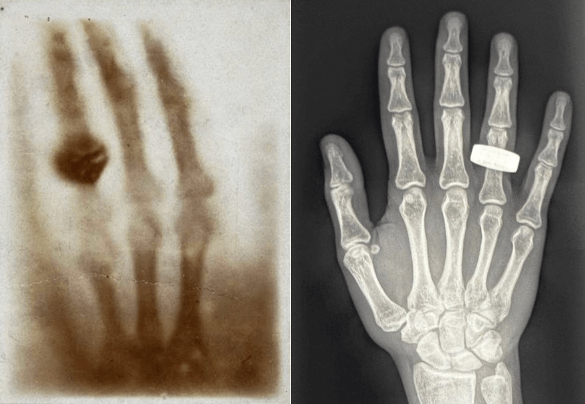
In 1895, Wilhelm Roentgen made history by capturing the first X-ray image of his wife’s hand, launching an entirely new era in medical diagnostics. Nearly 130 years later, history was made again. This time in orbit.
Earlier this month, NASA astronaut and flight engineer Dr. Satoshi Furukawa captured the first X-ray ever taken in space, aboard the International Space Station. Shared on Twitter/X, the image showcases not only technological advancement, but radiology’s growing relevance far beyond Earth.
This groundbreaking achievement, made possible with a compact, portable X-ray device proves that diagnostic imaging can be adapted to microgravity environments, opening the door to improved in-flight care and deep-space mission readiness.
A Quick Look Back: The Legacy of Wilhelm Roentgen
In 1895, German physicist Wilhelm Roentgen stumbled upon a mysterious form of radiation while experimenting with cathode rays. What he discovered, which he called X-rays, would forever change the course of medicine.
Roentgen’s first image, an X-ray of his wife’s hand revealing her bones and wedding ring, marked the first time physicians could see inside the human body without surgery. His discovery:
Remarkably, Roentgen refused to patent his work, believing it should serve the common good, a philosophy that still echoes across the radiology community today.
From a dimly lit lab in Germany to the vacuum of space, the spirit of Roentgen’s curiosity and innovation continues to shape the future of medicine.
Imaging in Microgravity: The First Medical X-ray Taken in Space
The new X-ray image was taken in microgravity, inside a four-person space capsule flying at orbital speeds of 17,500 miles per hour, about 200 miles above the Earth’s surface, documents Jennifer Chu from MIT News.1 The in-flight body scan was part of the SpaceXray project, one of 22 science experiments that astronauts conducted during the Fram2 mission.2
Dr. Lonnie Petersen, associate professor in MIT’s Department of Aeronautics and Astronautics and co-investigator on the SpaceXray project, explained the challenges of performing X-rays in space:
“To get hardware certified for spaceflight, it should be miniaturized and as lightweight as possible. There are also increased safety requirements because all devices work in a confined space. The increased requirements drive technology development.”
The project utilized a portable, specialized X-ray generator and detector developed by MinXray and KA Imaging, originally designed for battlefield use and adapted for spaceflight.3

The image on the left is Roentgen’s first X-ray image of his wife's hand; the image on the right is the first X-ray image taken in space.
Why This Moment Matters for Radiology
At Medality, we’re inspired by moments like this. They show us that radiology isn’t just about interpreting images; it’s about pushing boundaries, solving problems, and improving patient care everywhere. Even 200 miles above Earth.
Sources
1-3. MIT News. 3Q: MIT’s Lonnie Petersen on the first medical X-ray taken in space. Accessed April 28, 2025


