Cavernous Malformation
- Cavernous malformations (also known as cavernous angioma/hemangioma, cavernous venous malformation, or cavernoma) are the second most common intracranial vascular malformation; they are considered to be congenital
- They are slow-flow, low-pressure vascular malformations with no normal intervening brain parenchyma
- Most lesions are asymptomatic; seizures are the most common form of presentation when symptomatic; the risk of bleeding is 1-2% annually
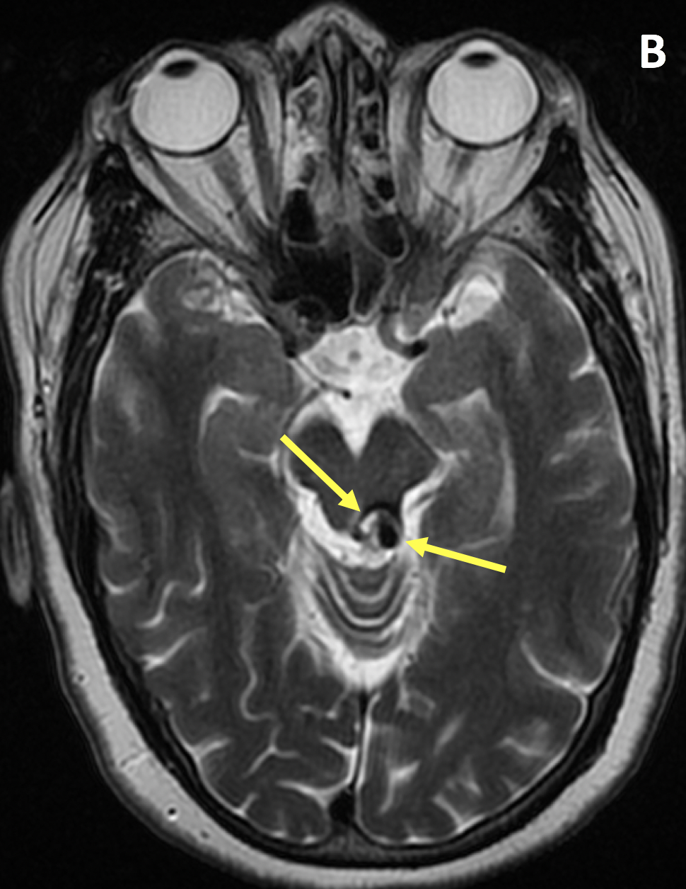
- Characteristic features on MRI include a reticulated pattern of mixed hyper- and hypointensity on T1- and T2-weighted imaging (so-called “popcorn” or “mulberry” appearance due to multiple episodes of hemorrhage), and a hypointense rim of hemosiderin surrounded by blooming artifact, best appreciated on T2-weighted or susceptibility-weighted imaging (SWI)
- If a recent bleed has occurred then surrounding edema may be present
- The lesions generally do not enhance, although enhancement is possible
- Multiple cavernous malformations can be seen when there is a familial component or in the setting of postradiation therapy
- T2-weighted SWI sequences, with their increased magnetic susceptibility effects, should always be performed during an evaluation for smaller or multiple lesions that may not be visible on fast spin-echo images
- Rigamonti D, Drayer BP, Johnson PC, Hadley MN, ZabramskiJ, Spetzler RF. The MRI appearance of cavernous malformations (angiomas). J Neurosurg 1987; 67(4):518-524
Case-based learning.
Perfected.
Try MRI Online Premium for free.
100+
Mastery Series video courses3000+
High-yield cases500+
Expert case reviewsUnlimited
CME & SA-CME creditsLearn from world renowned radiologists anytime,
actice on real, high-yield cases with MRI Online Premium.