Lateral Ankle Sprain
- 85% of ankle sprains are lateral, most commonly due to ankle inversion
- The lateral collateral ligament consists of the anterior talofibular ligament (ATFL), the calcaneofibular ligament (CFL), and the posterior talofibular ligament (PTFL)
- Lateral ankle sprains can be graded by the number of ligaments involved (Grade 1 – 1 ligament, usually ATFL; Grade 2 – 2 ligaments, usually ATFL and CFL; and Grade 3 – all 3 ligaments)
- Routine ankle MR imaging is performed in the axial, coronal, and sagittal planes; plantar flexion allows better visualization of the calcaneofibular ligament; sequences include T1 and T2; marrow abnormalities are best evaluated with STIR
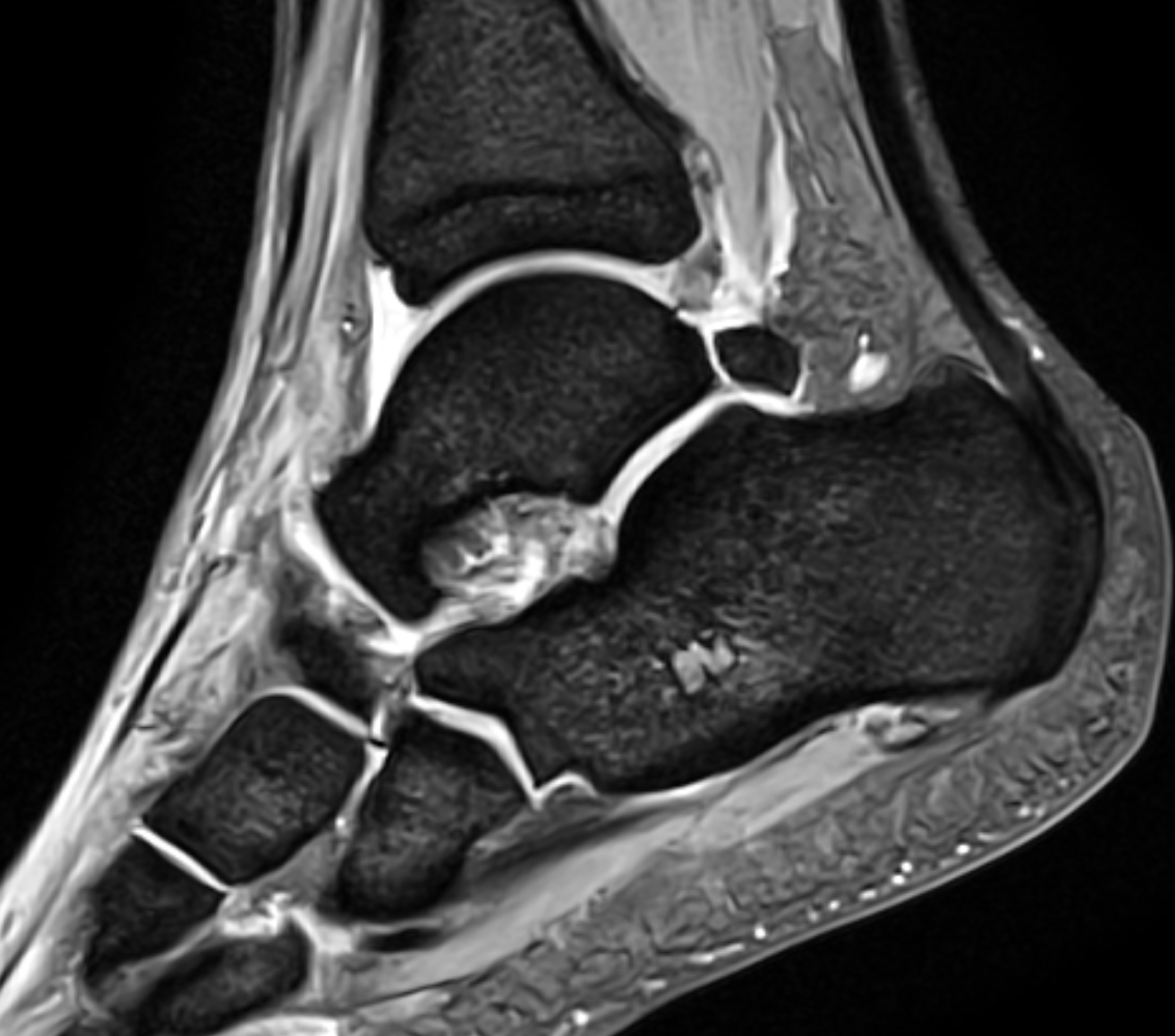
- Normal ligaments are thin, linear, low-signal-intensity structures joining adjacent bones, delineated by contiguous high-signal-intensity fat
- MRI of ligament injury shows discontinuity, detachment, thickening, thinning, or irregularity of the ligament
- Heterogeneity and increased intraligamentous signal intensity on FS or T2-weighted images is indicative of intrasubstance edema or hemorrhage
- Obliteration of the fat planes around the ligament, extravasation of joint fluid into the adjacent soft tissues, and talar contusions may also be seen
- Fluid within the peroneal tendon sheath can be a secondary sign of calcaneofibular ligament injury
- Chronic ligamentous tears manifest as thickening, thinning, elongation, and wavy or irregular contour of the ligament with no residual marrow or soft-tissue edema or hemorrhage
- Decreased signal intensity in the fat abutting the ligaments is indicative of scarring or synovial proliferation
- Perrich KD, Goodwin DW, Hecht PJ, Cheung Y. Ankle Ligaments on MRI: Appearance of Normal and Injured Ligaments. AJR 2009; 193:687–695
- Rosenberg ZS, Beltran J, Bencardino JT. Imaging of the Ankle and Foot. RadioGraphics 2000; 20:S153–S179
Case-based learning.
Perfected.
Try MRI Online Premium for free.
100+
Mastery Series video courses3000+
High-yield cases500+
Expert case reviewsUnlimited
CME & SA-CME creditsLearn from world renowned radiologists anytime,
actice on real, high-yield cases with MRI Online Premium.