Neurofibromatosis, Type 1 (NF1)
- NF1 is the most common neurocutaneous disorder (phakomatosis)
- It is an autosomal dominant mutation of the neurofibromin gene, which is a tumor suppressor gene of the RAS/MAPK pathway, referred to as a rasopathy
- The diagnosis of NF1 is clinical and criterion-based
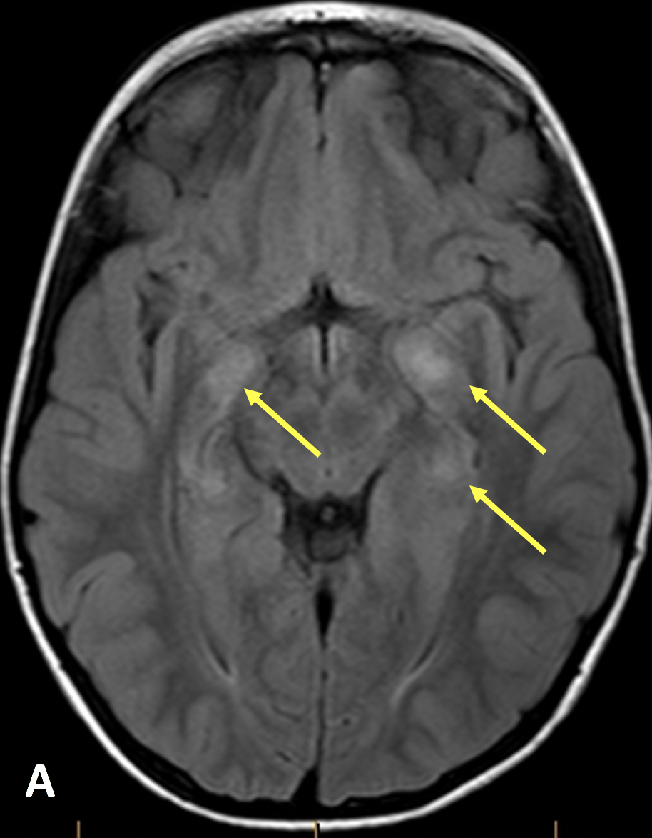
- On MRI, NF1 presents as areas of high signal on T2/FLAIR sequences in the deep white matter and basal ganglia; these areas represent myelin vacuolation that don’t enhance and often resolve during the second decade
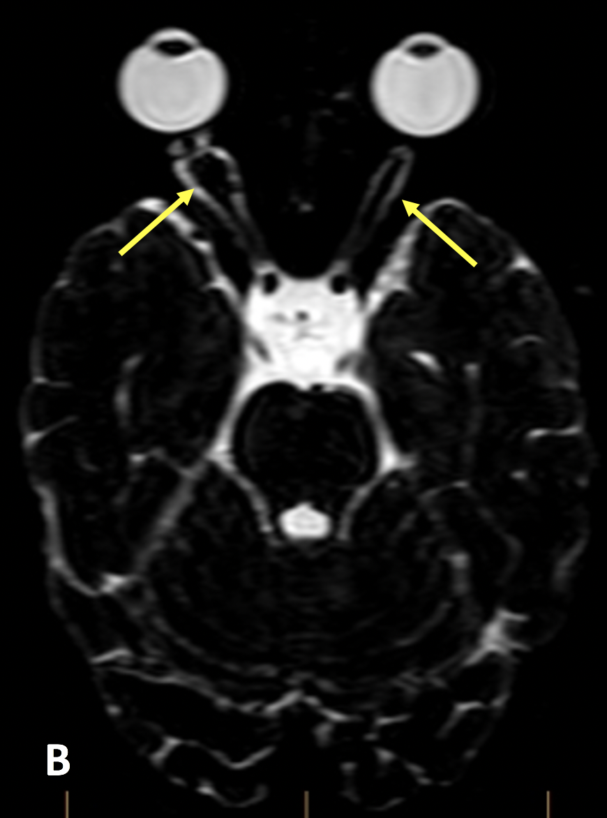
- Optic pathway gliomas are associated with NF1 and are best seen on thin-section fluid sensitive sequences (such as CISS or SPACE); they typically present as fusiform enlargement of parts of the optic pathways; they may enhance and be accompanied by tortuosity of the nerve and dilatation of the optic nerve sheath
- Buphthalmos (enlargement of the globe) can result from increased intraocular pressure
- Greater sphenoid wing dysplasia may be seen, best on CT, but also on MRI as dark signal cortical enlargement; this feature can result from neurofibromas or CSF pulsation
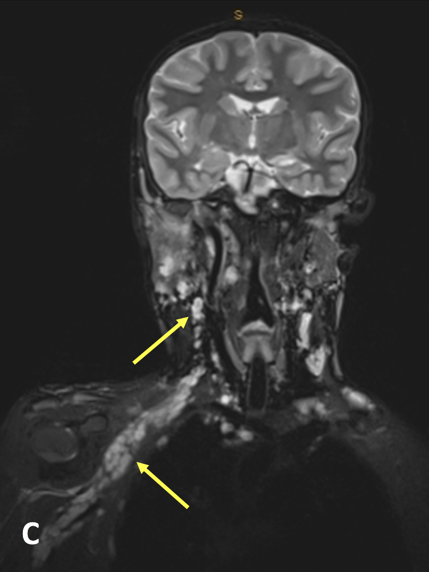
- Plexiform neurofibromas are STIR/T2 FS hyperintense fusiform and nodular masses along the course of nerves, often seen coalescing along the brachial, lumbar plexi and various cranial nerves
- Dural ectasia, a result of cerebrospinal fluid (CSF) pulsation, is seen on T2 sequences as outpouching of high signal CSF into the neural foramina and scalloping of the vertebral bodies
- Another manifestation of NF1 in the brain is moyamoya disease, which is caused by blocked arteries at the base of the brain; the name moyamoya means “puff of smoke” in Japanese and describes the appearance of tiny vessels that form to compensate for the blockage
- Plexiform neurofibromas may undergo malignant degeneration and surveillance imaging is performed to monitor for new enhancement or growth
- Ferner RE, Huson SM, Thomas N, et al. Guidelines for the diagnosis and management of individuals with neurofibromatosis 1. Journal of Medical Genetics 2007; 44(2):81-88
- Van Meerbeeck S, Verstraete K, Janssens S, Mortier G. Whole body MR imaging in neurofibromatosis type 1. European Journal of Radiology 2009; 69(2):236-242
Case-based learning.
Perfected.
Try MRI Online Premium for free.
100+
Mastery Series video courses3000+
High-yield cases500+
Expert case reviewsUnlimited
CME & SA-CME creditsLearn from world renowned radiologists anytime,
actice on real, high-yield cases with MRI Online Premium.