Optic Neuritis (ON)
- Optic neuritis (ON) is classically a demyelinating process of the optic nerve, presenting as orbital pain, decreased vision or dyschromatopsia (change in color perception) that progresses over several days
- Of patients presenting with ON, 40% are eventually diagnosed with multiple sclerosis
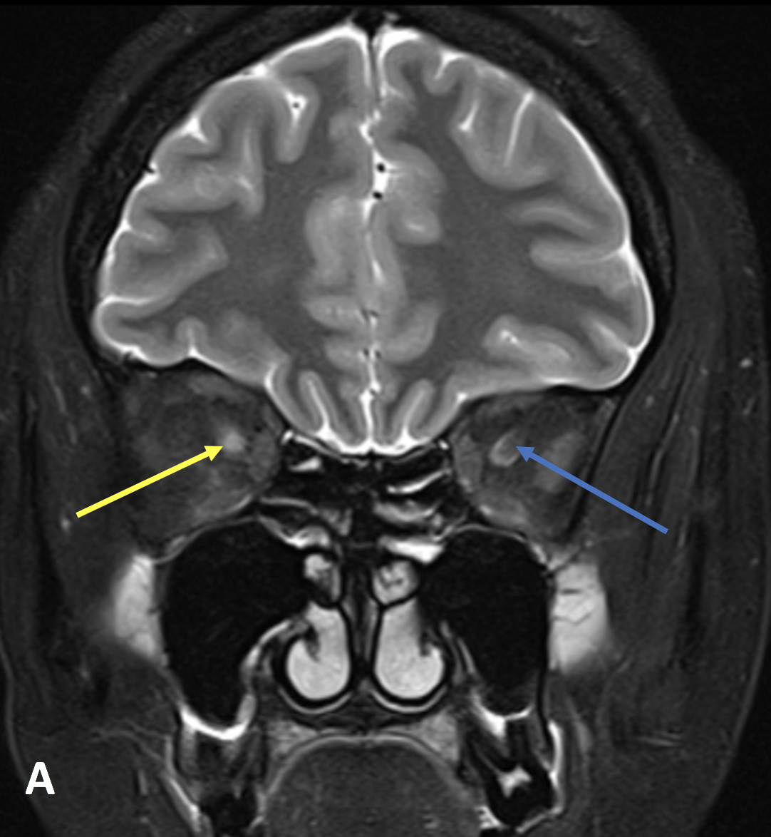
- In the setting of neuromyelitis optica (which affects the optic nerve and spinal cord), the ON changes are often bilateral
- On T2-weighted MR images, the optic nerve will be asymmetrically bright and may be mildly enlarged
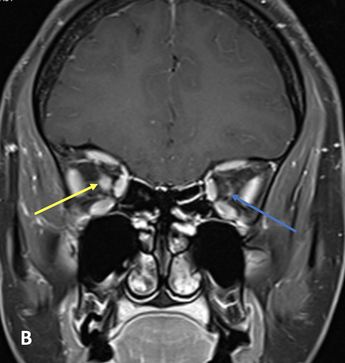
- T1 post-contrast imaging will show enhancement of the nerve
- In the chronic phase, the nerve will atrophy, best demonstrated on thin-section fluid sensitive sequences
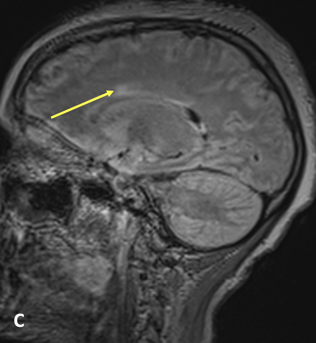
- In the presence of ON, it is important to evaluate the brain for lesions disseminated in space (i.e., separate lesions in the subcortical, juxtacortical, and infratentorial brain/spinal cord) and time (i.e., enhancing and nonenhancing lesions) according to the revised McDonald Criteria for multiple sclerosis
- Differential considerations for ON include optic nerve glioma (marked enlargement of the nerve) and optic nerve sheath meningioma (calcification and thickened enhancement of the nerve sheath but a normal nerve)
- Khanna S, Sharma A, Huecker J, Gordon M, Naismith RT, Van Stavern GP. Magnetic resonance imaging of optic neuritis in patients with neuromyelitis optica versus multiple sclerosis. Journal of Neuro-Ophthalmology 2012; 32(3):216-20
- Polman CH, Reingold SC, Banwell B, Clanet M, Cohen JA, Filippi M, Fujihara K, Havrdova E, Hutchinson M, Kappos L, Lublin FD. Diagnostic criteria for multiple sclerosis: 2010 revisions to the McDonald criteria. Annals of Neurology 201; 69(2):292-302
Case-based learning.
Perfected.
Try MRI Online Premium for free.
100+
Mastery Series video courses3000+
High-yield cases500+
Expert case reviewsUnlimited
CME & SA-CME creditsLearn from world renowned radiologists anytime,
actice on real, high-yield cases with MRI Online Premium.