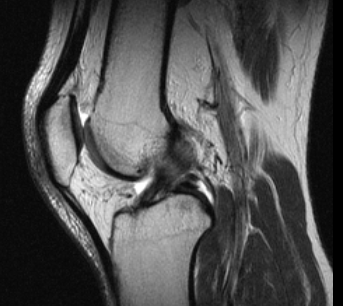
CASE

Knee – PCL tear

CASE HISTORY

28-year-old male with pain tightness and limited extension with some swelling since fall and twisting injury 3 weeks ago.

TECHNICAL FACTORS

Long- and short-axis fat- and water-weighted images were performed on a 0.95-Tesla. 

KEY IMAGES

CASE FINDINGS

  • Extensor tendons are intact
  • Moderate-sized hemarthrosis
  • High-grade tear of the proximal anterior cruciate ligament
  • Diffuse prepatellar and pretibial contusion versus closed degloving injury (Morel-Lavallee syndrome)
  • Minimally depressed comminuted posteromedial tibial plateau fracture with 3mm of depression. Extensive nondisplaced fracture in the posterolateral tibial plateau
  • Tibiofibular capsular sprain. Likely sprain of the arcuate ligament
  • Grade 2 sprain with interstitial PCL tear
  • Contusion in the posterior root of the lateral meniscus. No traumatic meniscal tears are demonstrated
  • Grade 1 to 2 sprain of the tibial collateral ligament. Lateral collateral complex low-grade sprain noted. Popliteus myotendinous junction sprain. Tendon is intact
  • Popliteal neurovascular structures are unremarkable. Chondral contusion demonstrated in the lateral femoral condyle
  • Low-grade patellofemoral chondromalacia
  • Tiny peripheral vertical meniscus red red zone body microtear. Possible knee subluxation.

CASE CONCLUSION

  • Pivot-shift injury with high-grade ACL tear nondisplaced fracture of the posterolateral tibial plateau grade 2 tibial collateral ligament sprain chondral contusion laterally and contusion of the posterior root of the lateral meniscus without traumatic tear. Interstitial PCL tear. Diffuse capsular sprain. 
  • Contrecoup injury with mildly depressed fracture of the posteromedial tibial plateau and low-grade fibular collateral sprain.
  • Grade 2 sprain with partial tear of the posterior cruciate ligament.
  • Posterolateral corner injury with popliteus myotendinous junction sprain (tendon otherwise intact). Tibiofibular capsular and likely arcuate ligament sprain. 
  • Diffuse swelling in the anterior superficial soft tissues as described.
  • Tiny peripheral vertical meniscus red red zone body microtear. Possible knee subluxation?

Case-based learning.
Perfected.

Learn from world renowned radiologists anytime, anywhere and practice on real, high-yield cases with Medality membership.

  • 100+ Mastery Series video courses
  • 4,000+ High-yield cases with fully scrollable DICOMs
  • 500+ Expert case reviews
  • Unlimited CME & CPD hours

Related Cases

Knee – Bucket handle meniscal tear

Read More

Knee – ACL tear

Read More

Knee – PCL tear

Read More

Case-based learning.
Perfected.

Try MRI Online Premium for free.

Customers served! 0 +  Mastery Series video courses
Customers served! 0 +  High-yield cases
Customers served! 0 +  Expert case reviews

Unlimited

CME & SA-CME credits

Learn from world renowned radiologists anytime,
practice on real, high-yield cases with MRI Online Premium.