CASE HISTORY
- 19-year-old male with soccer accident with swelling grinding and medial knee pain. Prior arthroscopic ACL reconstruction surgery using bone-patellar tendon-bone allograft
TECHNICAL FACTORS
- Long- and short-axis fat- and water-weighted images were performed
CASE FINDINGS
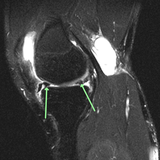
- Patient is status post ACL reconstruction surgery. The ACL graft is unusually vertical in configuration. Additionally the proximal aspect of the ACL graft (adjacent to the femoral tunnel) is attenuated/thinned in comparison to the remainder of the ACL graft compatible with graft failure deficiency and tear.
- A bucket-handle tear of the medial meniscus is present with a displaced bucket-handle fragment within the medial aspect of the intercondylar notch. Intermeniscal ligament vertical tear of posterior rim. This is new since the prior MRI examination. No lateral meniscal tear is present.
- A small focal area of high grade (grade 3 to 4) chondromalacia with mild underlying subchondral fibrocystic change is present within the medial compartment involving the posterior aspect of the weightbearing medial femoral condyle and overlying the posterior horn of the medial meniscus (sagittal T2 fat-sat series 10 image 23). This measures approximately 5mm in diameter and is best appreciated on sagittal T2 fat-sat and proton density series 10 and 9 image 23.
- No chondromalacia is apparent within the lateral or patellofemoral compartments.
- A large knee effusion is present. A moderate-sized Baker’s cyst is present within the posteromedial aspect of the knee with a small amount of fluid tracking inferiorly from the Baker’s cyst along the medial head of the gastrocnemius muscle compatible with partial cyst dehiscence/rupture.
- The PCL medial and lateral collateral ligaments and extensor mechanism are intact.
CASE CONCLUSION
- A bucket-handle tear of the medial meniscus is present with a displaced medial meniscal bucket-handle fragment within the medial aspect of the intercondylar notch. Intermeniscal ligament vertical tear of posterior rim
- Patient is status post ACL reconstruction surgery. The ACL graft has an unusually vertical configuration. Additionally the proximal aspect of the ACL graft (adjacent to the femoral tunnel) is attenuated/thinned compatible with graft failure deficiency
- Small approximately 5mm focal area of high grade chondromalacia within the posterior aspect of the weightbearing medial femoral condyle overlying the posterior horn of the medial meniscus with mild underlying subchondral fibrocystic change
- Large knee effusion
- Moderate-sized partially dehisced Baker’s cyst
Case-based learning.
Perfected.
Learn from world renowned radiologists anytime, anywhere and practice on real, high-yield cases with Medality membership.
- 100+ Mastery Series video courses
- 4,000+ High-yield cases with fully scrollable DICOMs
- 500+ Expert case reviews
- Unlimited CME & CPD hours


