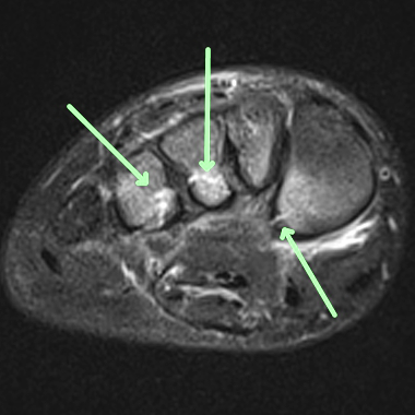
CASE HISTORY
- 1st and 3rd metatarsal fractures.
TECHNICAL FACTORS
- Long- and short-axis fat- and water-weighted images were performed.
CASE FINDINGS
- Transverse fracture at the base of the 4th comminuted fracture at the base of the 3rd nondisplaced microtrabecular or macrotrabecular fracture at the base of the 2nd microtrabecular injury of the epiphysis of the base of the 1st and microtrabecular injury of the navicular are noted.
- Other areas of microtrabecular injury also include an anterolateral calcaneus cuboid and cuneiforms.
- The Lisfranc ligament is torn.
- The C1-M3 ligament is torn. The C1-M2 ligament although not well seen may also be torn.
- No divergence at the Lisfranc articulation is noted; however to ascertain the nature and position of multiple fracture fragments thin-section CT with reformatting is strongly urged.
CASE CONCLUSION
- Midfoot fracture-subluxation injury with Lisfranc C1-M3 and possibly C1-M2 ligament tears
Case-based learning.
Perfected.
Learn from world renowned radiologists anytime, anywhere and practice on real, high-yield cases with Medality membership.
- 100+ Mastery Series video courses
- 4,000+ High-yield cases with fully scrollable DICOMs
- 500+ Expert case reviews
- Unlimited CME & CPD hours


