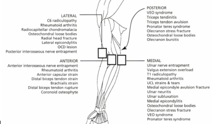
Diagnosis Definition
- Most biceps tendon ruptures are proximal at the shoulder; distal biceps tendon tears (DBTT) at the elbow account for 3-5% of biceps injuries
- DBTT can occur in football players and weightlifters, at or near the tendon’s insertion onto the radial tuberosity resulting from forced hyperextension of flexed and supinated arm
Imaging Findings
- MR imaging is performed in the axial plane with the arm extended, and in the longitudinal plane with the patient lying prone with the arm overhead, the elbow flexed to 90°, and the forearm supinated, so that the thumb points superiorly (FABS—flexed elbow, abducted shoulder, forearm supinated)
- In complete tears, MRI shows discontinuity with or without retraction (best seen on longitudinal FABS); the proximal tendon is enlarged with high signal
- Partial tears are seen as an increase in caliber and abnormal contour with high intratendinous signal
- High signal T2 peritendinous fluid (edema, bursitis, or hemorrhage) may be seen
Pearls
- If the bicipital aponeurosis is intact (best seen on axial view), there may be no tendon retraction
References
- Alentorn-Geli E, et al. Distal biceps tendon injuries: a clinically relevant current concepts review. EFORT Open Reviews 2016; 1:316-324.
- Chew ML, Giuffre BM. Disorders of the distal biceps brachii tendon. RadioGraphics 2005; 25:1227-1237.
- Pomeranz SJ, et al. Category B: Elbow. Gamuts & Pearls in MRI & Orthopedics. 1997; P. 60
- Sonin AH, et al. MR Imaging of Sports Injuries in the Adult Elbow. American Journal of Roentgenology 1996; 167(327):325-331.
Case-based learning.
Perfected.
Learn from world renowned radiologists anytime, anywhere and practice on real, high-yield cases with Medality membership.
- 100+ Mastery Series video courses
- 4,000+ High-yield cases with fully scrollable DICOMs
- 500+ Expert case reviews
- Unlimited CME & CPD hours

