Diagnosis Definition
- A SDH collects between the inner layer of the dura and the arachnoid
- The typical SDH occurs in infants, young adults in motor vehicle accidents, individuals who suffer from chronic alcoholism, people on anticoagulation drugs, or the elderly (due to increased space between the brain and skull, often compounded by frequent falls)
- The bleeding source is often from tearing of bridging cortical veins that cross the subdural space to enter a dural venous sinus
- Unlike epidural hematoma (EDH), SDH can occur from relatively minor head trauma, often without associated fractures
Imaging Findings
- Depending on whether acute, subacute, or chronic, SDH will have a variable appearance on MRI
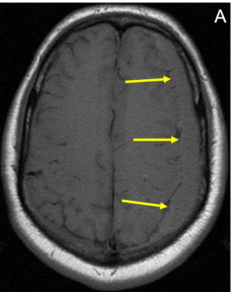
- SDHs are usually located over the cerebral convexities, are crescentic or curvilinear in shape, and are mostly limited by dural reflections but not by sutures
- FLAIR imaging can be most useful in delineating extra axial fluid collections, showing them as hyperintense to CSF in all phases of chronicity
KEY IMAGES

Pearls
- Since they are not limited by sutures, SDHs can be quite elongated in the anteroposterior dimension, distinguishing them from EDHs, which are biconvex or lentiform in shape and limited by sutures
- Posteriorly, subdural blood can outline the superior sagittal sinus, resulting in a “pseudodelta” appearance
- SDHs of different ages in infants and young children may be a sign of non-accidental trauma (e.g., child abuse)
References
- Atkinson JL, Lane JI, Aksamit AJ. MRI depiction of chronic intradural (subdural) hematoma in evolution. J Magn Reson Imaging 2003; 17(4):484-6
Case-based learning.
Perfected.
Learn from world renowned radiologists anytime, anywhere and practice on real, high-yield cases with Medality membership.
