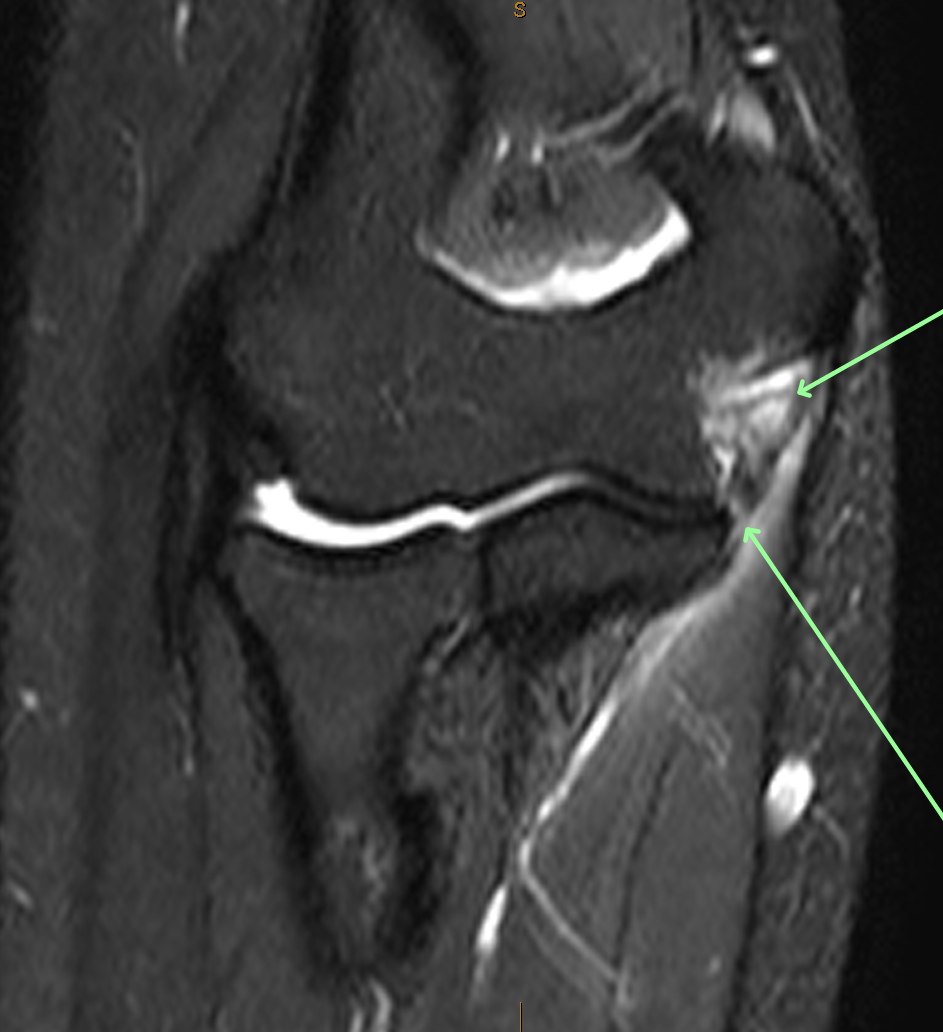
Diagnosis

Ulnar Collateral Ligament (UCL) tear

Diagnosis Definition

  • The ulnar collateral ligament (UCL) originates from the medial humeral epicondyle and inserts on the medial proximal ulna
    UCL tears result from chronic repetitive trauma (baseball pitching) or an acute fall on an outstretched hand
  • Most tears are complete; partial tears most commonly occur along the humeral attachment in baseball pitchers
  • The ulnar collateral ligament (UCL) originates from the medial humeral epicondyle and inserts on the medial proximal ulna

Imaging Findings

  • Routine elbow MRI sequences include sagittal PD, and axial and coronal T1 and T2 Fat-Sat (FS); MR arthrography (MRA) sequences include axial T1, axial T2 FS, coronal T1 FS, coronal T2 FS, and sagittal T2 FS after injecting the elbow joint with contrast
  • UCL tears on MRI are seen as T2 signal hyperintensity, swelling, and UCL irregularity/poor definition, abnormal laxity, or discontinuity
  • Heterotopic ossification may be seen in chronic tears

KEY IMAGES

Pearls

  • A partial tear of the UCL on MRA is seen as intra-articular contrast extension distally from the joint line along the cortical margin of the sublime tubercle referred to as the “T-sign”
  • A few mm of separation of the distal insertion of the anterior bundle of the UCLand the sublime tubercle may be seen in approximately 50% of normal individuals
  • Periligamentous edema involving the adjacent flexor digitorum superficialis muscle may be the only sign of low grade UCL injury

References

1.Delgado J, Jaramillo D, Chauvin NA. Imaging the Injured Pediatric Athlete: Upper Extremity. RadioGraphics 2016; 36(6): 1672-1687

2. Bucknor MD, Stevens KJ, Steinbach LS. Elbow Imaging in Sport: Sports Imaging Series. Radiology 2016; 279(1):12-28

Case-based learning.
Perfected.

Learn from world renowned radiologists anytime, anywhere and practice on real, high-yield cases with Medality membership.

  • 100+ Mastery Series video courses
  • 4,000+ High-yield cases with fully scrollable DICOMs
  • 500+ Expert case reviews
  • Unlimited CME & CPD hours

Related Diagnosis

Neurofibromatosis, Type 1 (NF1)

Read More

Inverted Papilloma (IP)

Read More

Optic Neuritis (ON)

Read More

Case-based learning.
Perfected.

Try MRI Online Premium for free.

Customers served! 0 +  Mastery Series video courses
Customers served! 0 +  High-yield cases
Customers served! 0 +  Expert case reviews

Unlimited

CME & SA-CME credits

Learn from world renowned radiologists anytime,
practice on real, high-yield cases with MRI Online Premium.