CASE HISTORY
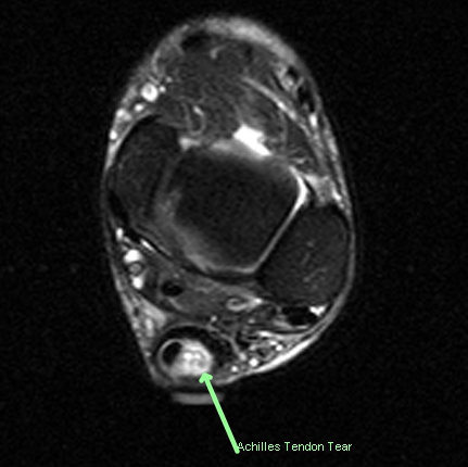
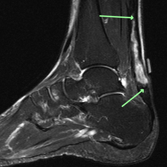
- Achilles injury or rupture evaluate for length and extent.
TECHNICAL FACTORS
- Long- and short-axis fat- and water-weighted images were performed
CASE FINDINGS
- Tendinosis within an enlarged Achilles is associated with a massive rupture that is encased in either: a) The peripheral fibers of the Achilles as a thin rim. b) The paratenon. c) Fibrous tissue.
- The length of the tear is measured by MRI at 7cm and tapers proximally.
- The tear begins 2cm above the superior calcaneal protuberance.
- Incidentally noted is small ankle effusion and mild to moderate midfoot arthrosis with subcortical erosions and pseudocysts.
- Mild calcaneal periostitis and small inferior spur.
CASE CONCLUSION
- 7cm long tear of the Achilles encased in a hypointense rim whose differential diagnosis is given and with a small tubular structure within most likely representing a well-developed residual persistent or untorn plantaris. See series 401 image 19 as an ex
Case-based learning.
Perfected.
Learn from world renowned radiologists anytime, anywhere and practice on real, high-yield cases with Medality membership.
- 100+ Mastery Series video courses
- 4,000+ High-yield cases with fully scrollable DICOMs
- 500+ Expert case reviews
- Unlimited CME & CPD hours


