CASE HISTORY
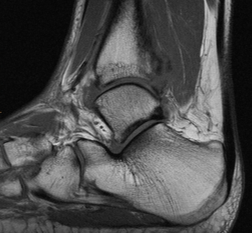
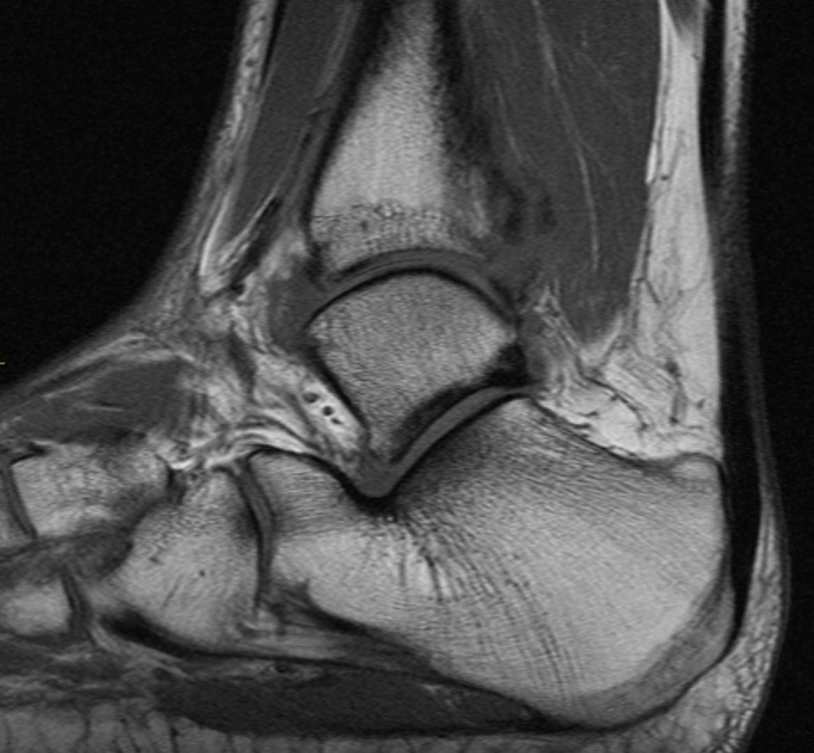
- Evaluate for talar dome OCD in a patient with syndesmosis widening.
TECHNICAL FACTORS
- Long- and short-axis fat- and water-weighted images were performed.
CASE FINDINGS
- High ankle intercalary sprain/rupture involves the anterior tibiofibular ligament the syndesmotic membrane and the posterior tibiofibular ligament. Only the upper fibers of the anterior talofibular ligament are involved and are swollen. The deltoid is swollen but remains intact. The typical bone injury e.g. microtrabecular fracture and periosteal hemorrhage of the posterior malleolus is present as is often the case with high-grade high ankle sprains. A pattern of posterior malleolar fragmentation might be ascertained with thin section CT if so desired
- Hemarthrosis with coagulated blood in the anterior capsular recess.
- No signs of OCD or loose body are noted.
- The transverse tib/fib ligament and remainder of the crural system are likely intact.
CASE CONCLUSION
- High ankle sprain intercalary variety involving anterior syndesmotic and posterior components with associated Volkmann fragment. No talar dome OCD
Case-based learning.
Perfected.
Learn from world renowned radiologists anytime, anywhere and practice on real, high-yield cases with Medality membership.
- 100+ Mastery Series video courses
- 4,000+ High-yield cases with fully scrollable DICOMs
- 500+ Expert case reviews
- Unlimited CME & CPD hours


