CASE HISTORY
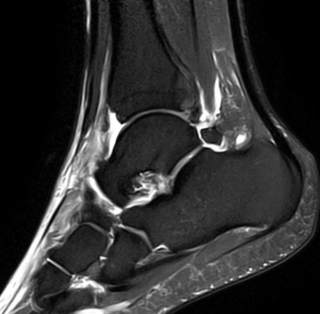
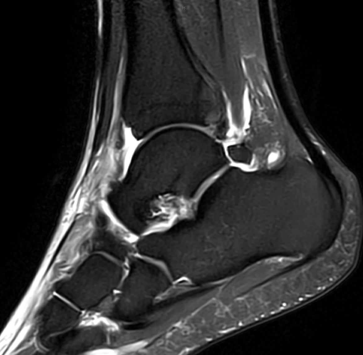
- Stepped on another player’s foot and felt a pop in the ankle
TECHNICAL FACTORS
- Long- and short-axis fat- and water-weighted images were performed. 1.5T High Field Oval
CASE FINDINGS
- Effusion and hemarthrosis.
- Anterior talofibular ligament ruptured.
- Calcaneofibular ligament ruptured.
- Posterior talofibular ligament swollen but intact.
- High ankle and syndesmosis normal.
- Peroneal retinaculum torn.
- Peroneus longus and brevis normal.
- Medial tendon group normal.
- Anterior tendon group normal.
- Posterior tendon group normal.
- Subtalar ligaments abnormal:
- a. Talocalcaneal interosseous ligament intact.
- b. Cervical ligament ruptured.
- c. Inferolateral retinaculum or stem ligament torn.
- Skeleton: No fractures no OCDs and large os trigonum.
- Soft Tissues: Large anterolateral hematoma with soft tissue swelling
CASE CONCLUSION
- Two part ankle sprain/rupture low ankle. High ankle spared. Hematoma. Subtalar ligamentous injury involving cervical and inferolateral ligamentous complex
Case-based learning.
Perfected.
Learn from world renowned radiologists anytime, anywhere and practice on real, high-yield cases with Medality membership.
- 100+ Mastery Series video courses
- 4,000+ High-yield cases with fully scrollable DICOMs
- 500+ Expert case reviews
- Unlimited CME & CPD hours


