CASE

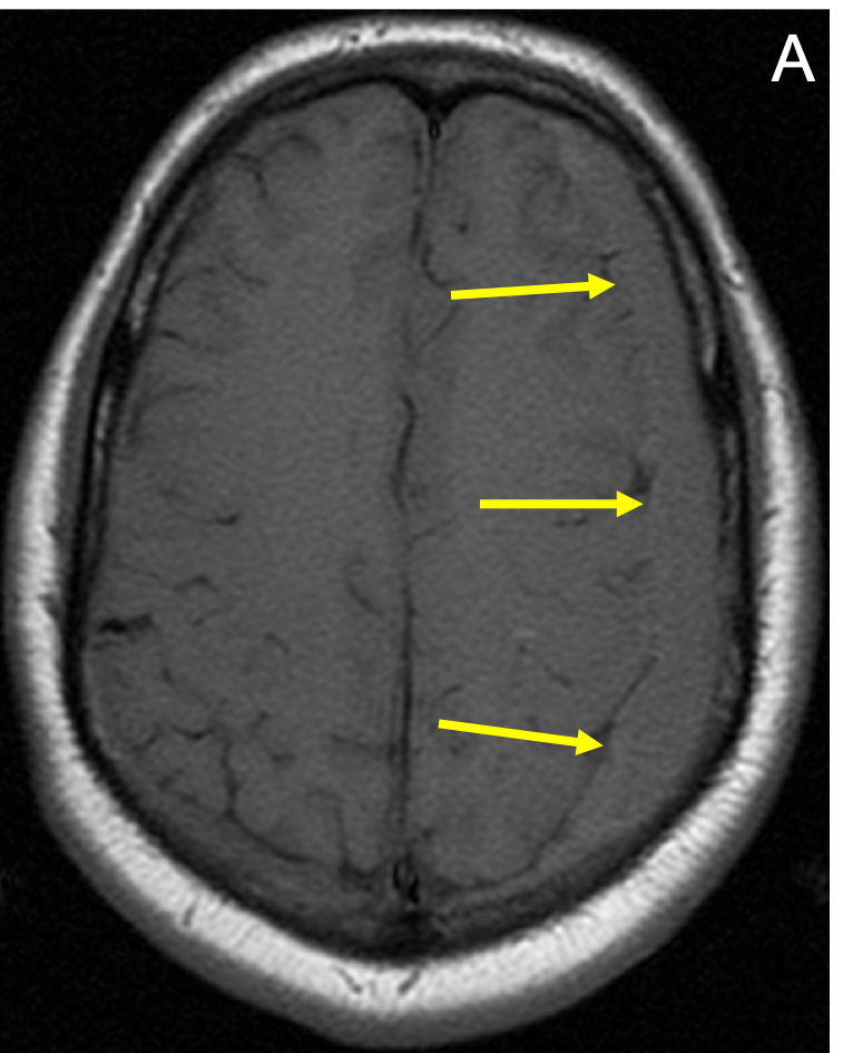
Brain – Subdural Hematoma (SDH)

Chronic SDH in a 64-yea-old man with a headache. T1-weighted (A) and T2-weighted (B) axial MRI images show an elongated, extra axial, frontoparietal, crescentic fluid collection (arrows) that is hyperintense to brain.

CASE HISTORY

22-year-old male with long-standing history of headache. Extraaxial fluid collection on left.

TECHNICAL FACTORS

Long- and short-axis fat- and water-weighted images were obtained before and after contrast administration. 10cc of OptiMARK injected. 

KEY IMAGES

Chronic SDH in a 64-yea-old man with a headache. T1-weighted (A) and T2-weighted (B) axial MRI images show an elongated, extra axial, frontoparietal, crescentic fluid collection (arrows) that is hyperintense to brain.Chronic SDH in a 64-yea-old man with a headache. T1-weighted (A) and T2-weighted (B) axial MRI images show an elongated, extra axial, frontoparietal, crescentic fluid collection (arrows) that is hyperintense to brain.

CASE FINDINGS

  • Inflammatory pseudotumor is seen at the level of the dens. Nominal effacement anterior aspect of the upper cervicomedullary junction. Sagging of the tonsils and medulla within the posterior fossa and effacement of the prepontine cistern noted. 
  • Mixed predominantly T2 dark marginally hyperintense subdural hematoma left frontoparietal zone with oblique coronal thickness 11mm. No appreciable shift. Mild effacement of the ipsilateral lateral ventricle and left temporal horn. Diffuse signal hyperintensity within the right extraaxial space and diffuse exaggerated concentric supratentorial dural thickening follows contrast administration. No evidence of midline meningioma (see head CT report) 
  • Scattered moderate random patchy foci of white matter FLAIR and T2 hyperintensity predominantly in the frontal territory also demonstrated as sequela of metabolic or small vessel ischemic change. 
  • Partially empty sella. 
  • Expected flow voids are patent. 
  • No intraaxial region of abnormal enhancement. 
  • Mild ethmoid air cell mucosal thickening. Bilateral maxillary sinus mucous retention cysts. Left mucous retention cyst likely odontogenic in origin. 
     

CASE CONCLUSION

  • Suspect primary intracranial hypotension with exaggerated pachymeningeal enhancement sagging of the posterior fossa medulla and tonsils with precontrast right hemispheric marginal FLAIR hyperintensity. 
  • Mixed-signal predominantly chronic 11 mm diameter left frontoparietal chronic subdural collection or proteinaceous effusion from leak with mixed signal intensity. No associated shift or advanced sulcal effacement. 
  • Mild periventricular ependymitis granularis and scattered areas of small vessel arteriopathy and/or venopathy induced gliosis or leukoaraiosis are noted most often seen as manifestation of hypertension arteriosclerosis and/or metabolic factors such as hypercholesterolemia or hypertriglyceridemia. 
  • Consider total spine MRI to look for source of CSF leak which could be spinal or intracranial. Strongly consider removal of extra-axial collection. 
  • Effaced left ventricle due to subdural and dilated right ventricle system and temporal horn. Most likely hypotension with dural leak explains subdural as one unifying diagnosis. 
  • However sulcal effacement slightly enlarged ventricles raise the outside possibility of a combined issue of both chronic communicating hydrocephalus with a chronic dural leak. With subdural a spinal tap at this juncture may be contraindicated for pressures. 
     

Case-based learning.
Perfected.

Learn from world renowned radiologists anytime, anywhere and practice on real, high-yield cases with Medality membership.

  • 100+ Mastery Series video courses
  • 4,000+ High-yield cases with fully scrollable DICOMs
  • 500+ Expert case reviews
  • Unlimited CME & CPD hours

Related Cases

Knee – Bucket handle meniscal tear

Read More

Knee – ACL tear

Read More

Knee – PCL tear

Read More

Case-based learning.
Perfected.

Try MRI Online Premium for free.

Customers served! 0 +  Mastery Series video courses
Customers served! 0 +  High-yield cases
Customers served! 0 +  Expert case reviews

Unlimited

CME & SA-CME credits

Learn from world renowned radiologists anytime,
practice on real, high-yield cases with MRI Online Premium.