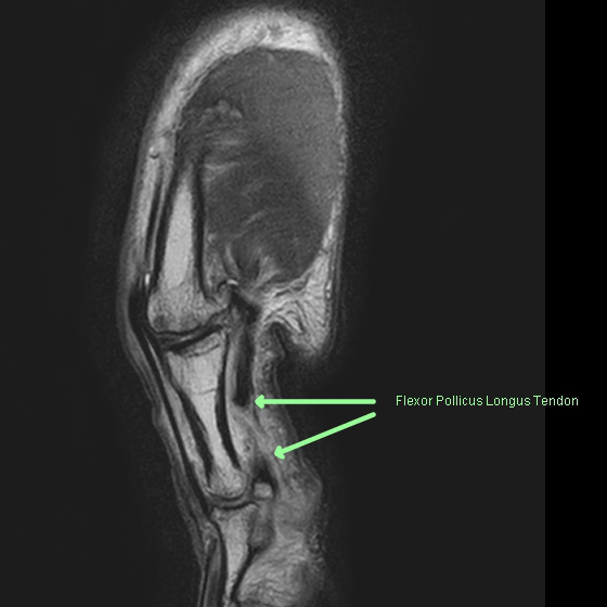
Diagnosis Definition
- The Achilles tendon is the strongest and thickest tendon in the body, connecting gastrocnemius and soleus calf muscles to the calcaneus
- Predisposing conditions include rheumatoid arthritis, systemic lupus erythematosus, diabetes mellitus, and gout
- Injuries include tendinitis, partial tear, complete tear, and chronic tear; generally during athletic activity
Imaging Findings
- Sequences include sagittal T1 and STIR, axial PD and T2 FS, and coronal PD and T2 FS
- The Achilles tendon is tapered, with uniformly dark signal on all sequences; in cross section, it has a C-shape that is concave toward the tibia, but becomes convex and thickened with tendinopathy or tears
- Partial tears are seen as thickening of the tendon with longitudinal bands of increased T1 and T2 signal
- In complete tears, MR shows tendon discontinuities with high T2 signal; tendon-end retraction may be seen
- With chronic tears, retracted tendon edges, and atrophy of tendon fibers and muscles are seen
KEY IMAGES

Pearls
- Because the Achilles tendon does not have a tendon sheath, tenosynovitis and fluid around the tendon are not seen; however, soft tissue edema in the soft tissues surrounding the tendon may be present after an acute tear
- Average Achilles tendon thickness is 6 mm
- Most tears occur 2-6 cm from insertion
References
- Schweitzer M, Karasick D. MR imaging of disorders of the Achilles tendon. American Journal of Roentgenology 2000; Vol. 175, Pp. 613-625.
Case-based learning.
Perfected.
Learn from world renowned radiologists anytime, anywhere and practice on real, high-yield cases with Medality membership.
