Diagnosis Definition
- Stroke occurs when decreased blood flow to the brain results in cell death (infarct/necrosis)
- There are two main types of stroke: ischemic (most common) due to lack of blood flow from thrombosis, embolism, systemic hypoperfusion, or cerebral venous sinus thrombosis, and hemorrhagic, due to bleeding
- Signs and symptoms are numerous and may include unilateral numbness or paralysis, problems understanding or speaking, dizziness, headache, or unilateral loss of vision
- Therapeutic options available in the first hours of the event are intravenous/intra-arterial thrombolysis or thrombectomy
Imaging Findings
- Non-contrast CT (NCCT) signs of ischemic stroke include low attenuation in the insula (insular ribbon sign) and basal ganglia (basal ganglia sign), hyperdense middle cerebral artery (representing intraluminal thrombus), blurring of the gray-white matter differentiation, hemorrhage, and sulcal effacement
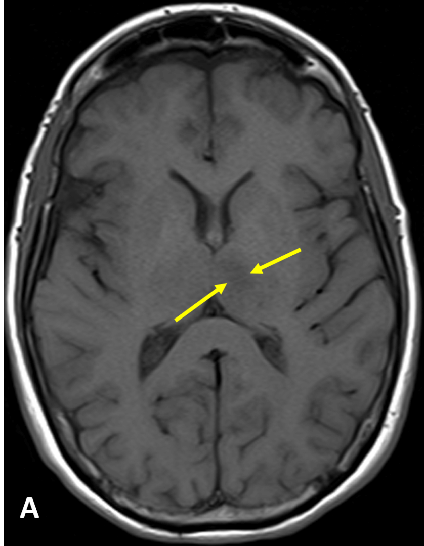
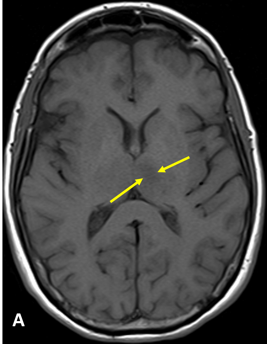
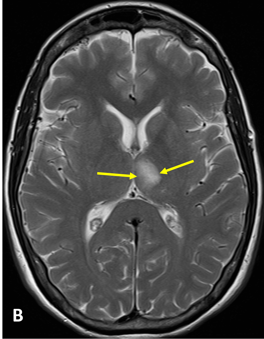
- The appearance of infarction on MRI depends on the stage: 1) hyperacute (< 24 hours) is isointense on T1 and iso- or hyperintense on T2/FLAIR, 2) early and late subacute (1-30 days) is hypointense on T1 and hyperintense on T2/FLAIR , and 3) chronic (> 1 month) is hypointense on T1 with volume loss/encephalomalacia and hypointense with surrounding hyperintensity on T2/FLAIR
- MRI shows restricted diffusion (e.g., high signal on DWI; initially decreased signal on ADC maps, gradually normalizing, then high signal after 10 days); DWI and ADC normalize after 30 days
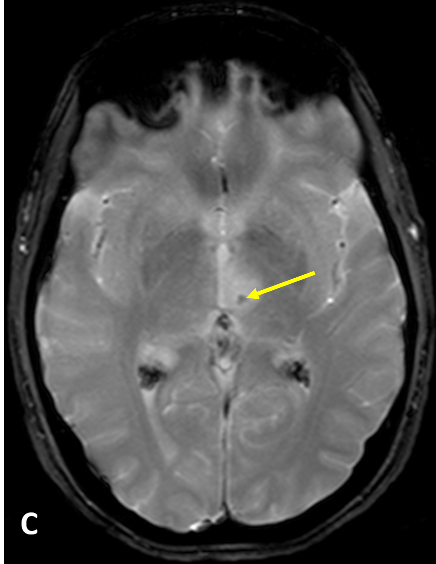
- Intraluminal thrombus and hemorrhage can be seen on MRI as blooming effect on gradient-echo (GRE) or susceptibility-weighted imaging (SWI)
- MRA/CTA can show clot and/or occluded blood vessels
Pearls
- FLAIR images demonstrate abnormal signal in the infarcted tissue approximately 6 hours after the event; thus, when the time of event is unknown, a mismatch between positive DWI and a negative FLAIR may suggest a time of onset of less than 6 hours
- Hemorrhagic transformation of ischemic stroke can result from reperfusion of ischemic-damaged endothelium and/or from thrombolytic therapy
References
- Kamalian S, Lev MH. Stroke imaging. Radiol Clin North Am 2019; 57(4):717-732
- Srinivasan A, Goyal M, et al. State of the art imaging of acute stroke. Radiographics 2006; 26:S75–S95
- Birenbaum D, Bancroft LW, Felsberg GJ. Imaging in acute stroke. West J Emerg Med 2011; 12(1):67-76
Case-based learning.
Perfected.
Learn from world renowned radiologists anytime, anywhere and practice on real, high-yield cases with Medality membership.
- 100+ Mastery Series video courses
- 4,000+ High-yield cases with fully scrollable DICOMs
- 500+ Expert case reviews
- Unlimited CME & CPD hours


