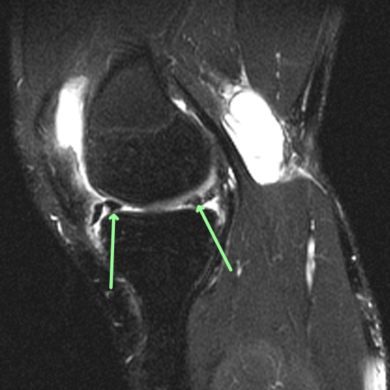
Diagnosis Definition
- Medial and lateral menisci are described as wedge-shaped or C-shaped pieces of cartilage sitting on the tibial plateaus that are tough and rubbery and help cushion and stabilize the knee joint
- Meniscal tears are among the most common knee injuries, especially in athletes
- Older people are prone to degenerative meniscal injuries
- Symptoms include pain, swelling, locked knee (or knee “giving way”), and limited range of motion
- Menisci have limited capacity for healing; surgical options include direct repair, trimming of unstable edges and fragments, and meniscectomy
Imaging Findings
- Routine knee MRI includes sagittal, coronal, and axial planes combined with fluid-sensitive fat-suppressed and non-fat-suppressed pulse sequences without contrast; field of view includes the suprapatellar recess at the superior extent and the proximal tibiofibular joint at the distal extent
- Each meniscus has an anterior horn, body, and posterior horn with no anatomical boundaries
- The primary finding of a tear is high signal within the normally dark meniscus that extends unequivocally to an articular surface
- Tears can be horizontal, longitudinal (also called circumferential or vertical), or radial
Pearls
- Horizontal cleavage tears are commonly degenerative and seen in older people
- Meniscal cysts may form at the periphery when joint fluid escapes through a meniscal tear
- A displaced longitudinal tear is a “bucket handle” tear
- Medial meniscus bucket handle tears can result in a double PCL sign
- Lateral meniscus bucket handle tears can produce the double anterior horn sign or double ACL sign
- Tears can be characterized by length, depth, shape, gap, displacement, stability, dysplasia (discoid)
References
- Rosas HG. Magnetic resonance imaging of the meniscus. Magn Reson Imaging Clin N Am 2014;22(4):493-516
- De Smet AA. How I diagnose meniscal tears on knee MRI. AJR Am J Roentgenol 2012;199(3):481-499
Case-based learning.
Perfected.
Learn from world renowned radiologists anytime, anywhere and practice on real, high-yield cases with Medality membership.
- 100+ Mastery Series video courses
- 4,000+ High-yield cases with fully scrollable DICOMs
- 500+ Expert case reviews
- Unlimited CME & CPD hours


