Diagnosis
Diagnosis Definition
- Cavernous malformation (CM) is a non-neoplastic venous malformation, which can occur sporadically, following radiation, or as a heritable condition
- It is a common incidental finding, but can become symptomatic due to hemorrhage
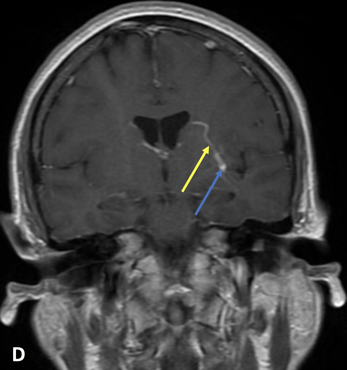
- The presence of an associated developmental venous anomaly (DVA), a congenital variant of cerebral venous drainage, increases the confidence of the diagnosis
Imaging Findings
-
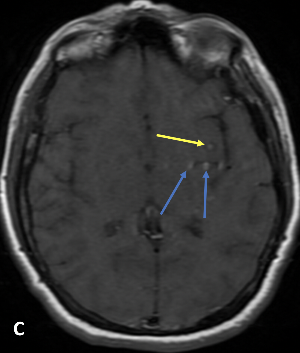
T2 weighted MRI sequences show a lobulated mass with a dark rim (known as “blooming artifact” and best seen on T2*); T1 and/or T2 bright signal may be seen centrally, depending on the age of the blood, creating a “popcorn lesion” because of the lobulated appearance
- Occasionally, an associated DVA can be seen as a collection of tiny flow voids coalescing into a vein directed towards a dural sinus or deep ependymal vein
- CT is neither sensitive nor specific for diagnosing CMs; if a CM is seen, it will usually appear hyperdense and somewhat ill-defined
- CMs are seldom seen on angiography
KEY IMAGES


Pearls
- CMs can become symptomatic when they bleed due to mass effect or local inflammation from blood products
- Resection is curative, although associated DVAs complicate resection because they result in a venous infarction
- Differential considerations include diffuse axonal injury (DAI), cerebral amyloid angiopathy, and vasculitis
References
- Dasgupta R, Fishman SJ. ISSVA classification. Seminars in Pediatric Surgery 2014; 23(4):158-161
- Starcevich J, Hart B, Bartlett M, Selwyn R, Morrison L, Hallstrom J. MRI sequence sensitivity for the detection of spinal cavernous malformations. 2016; available from https://digitalrepository.unm.edu/rad_pubs/8 on 10/17/2019
Case-based learning.
Perfected.
Learn from world renowned radiologists anytime, anywhere and practice on real, high-yield cases with Medality membership.
