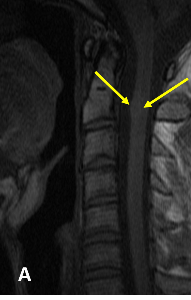
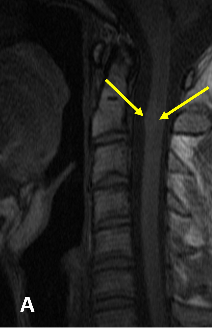
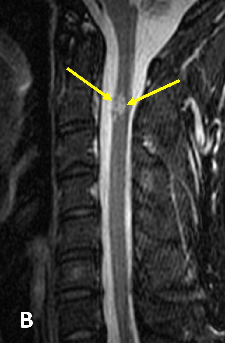
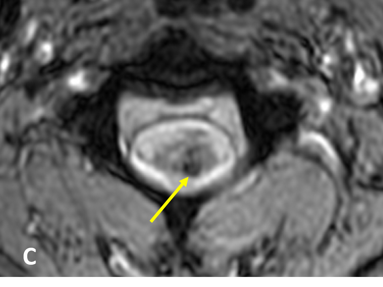
Diagnosis Definition
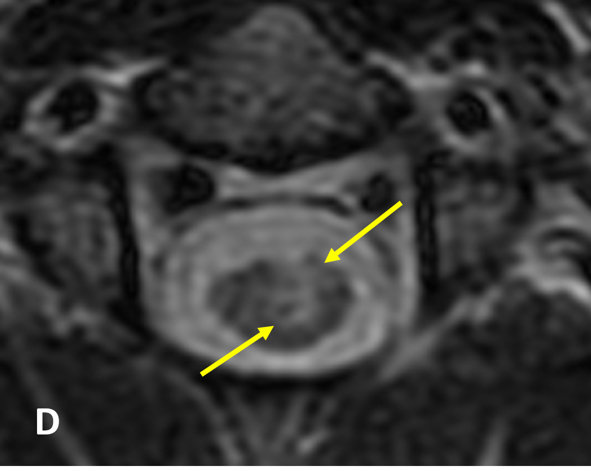
- Cavernous malformations (CMs, aka “cavernomas”) are slow flow vascular malformations with no intervening neuronal tissue; most are congenital
- Typically intramedullary and thoracic in location, spinal CMs represent ~5% of intramedullary lesions in adults
- Their propensity for internal hemorrhage can lead to progressive myelopathy related to mass effect
Imaging Findings
- Although the imaging appearance of CMs is variable, MRI typically shows a rounded mass that is heterogeneous in signal on T1-weighted images and predominantly hyperintense with a hypointense rim on T2 (described as having a “popcorn” or “mulberry” appearance)
- Almost all CMs have hypointense areas on GRE images and typically show minimal or no enhancement
- Extra- and intracellular methemoglobin and thrombosis account for the high-intensity signal on T1 and T2, while calcification, fibrosis, and acute and chronic blood products (e.g., hemosiderin) result in low-signal areas
- Cord expansion or edema is minimal unless there has been recent hemorrhage
Pearls
- Cavernous malformations can occur de novo after radiation therapy in the spinal cord as well as within the brain
- The risk of significant acute hemorrhage is low due to low venous pressure; however, recurrent hemorrhage within the confined intramedullary space can lead to severe neurological deterioration
References
- Rivera PP, Willinsky RA, Porter PJ. Intracranial cavernous malformations. Neuroimaging Clin N Am 2003; 13:27-40
- Jabbour P, Gault J, Murk SE, Awad IA. Multiple spinal cavernous malformations with atypical phenotype after prior irradiation: case report. Neurosurgery 2004; 55(6):1431
Case-based learning.
Perfected.
Learn from world renowned radiologists anytime, anywhere and practice on real, high-yield cases with Medality membership.
- 100+ Mastery Series video courses
- 4,000+ High-yield cases with fully scrollable DICOMs
- 500+ Expert case reviews
- Unlimited CME & CPD hours

