Diagnosis
Craniopharyngioma

Diagnosis
Craniopharyngioma is a slow-growing, rare, histologically benign brain tumor affecting children between the ages of 5 and 14 and older adults
It is extra-axial, arises near the pituitary gland, and commonly contains solid and cystic components
Pressure on the pituitary gland and optic nerves produces headache, visual symptoms and endocrine abnormalities
The most common type, adamantinomatous, is seen predominantly in children
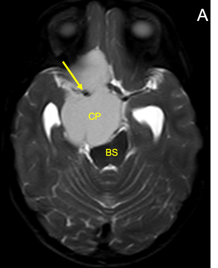
The tumor often contains single or multiple cysts filled with thick oily fluid rich in protein, blood products, and/or cholesterol, giving the so-called “motor oil” fluid appearance
Solid components are variable in intensity on T1 and T2 imaging and enhance vividly on T1 imaging with contrast
Cysts are variable in signal on T1 imaging due to high protein content and blood products, and variable but usually hyperintense on T2 imaging
Large tumors often compress the midbrain, causing obstructive hydrocephalus


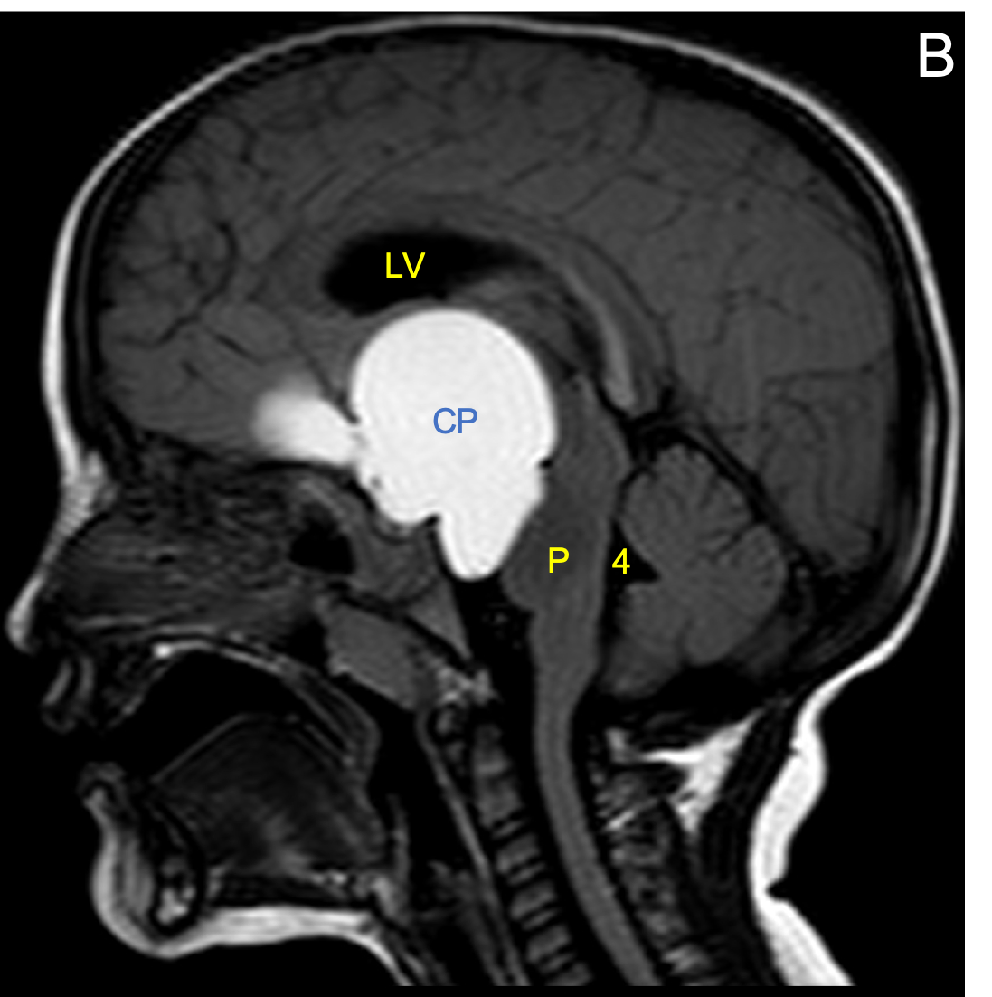
Appoximately 90% of craniopharyngiomas have calcifications, 90% have cysts, 90% enhance, and 90% are suprasellar
CT scanning is preferred over MRI for showing calcification in the tumor
Learn from world renowned radiologists anytime, anywhere and practice on real, high-yield cases with Medality membership.

Try MRI Online Premium for free.
Unlimited
CME & SA-CME credits
Learn from world renowned radiologists anytime,
practice on real, high-yield cases with MRI Online Premium.