Diagnosis
Diagnosis Definition
- Encephalomalacia (literally, “softening of the brain”) is a nonspecific term for the end result of liquefactive necrosis of brain parenchyma; it can be focal or diffuse, and can be seen in adults, children and even in utero
- Encephalomalacia can occur anywhere in the brain; after trauma, characteristic locations are anteroinferior frontal and temporal lobes
- The most common causes are hemorrhage or infarction; other causes include trauma (including surgery), neoplasm, infection, inflammation, and numerous other insults to the brain
- Leukoencephalomalacia refers to encephalomalacia of the white matter
- Areas of encephalomalacia are often surrounded by a rim of gliosis, which is the proliferation or hypertrophy of glial cells in response to injury
Imaging Findings
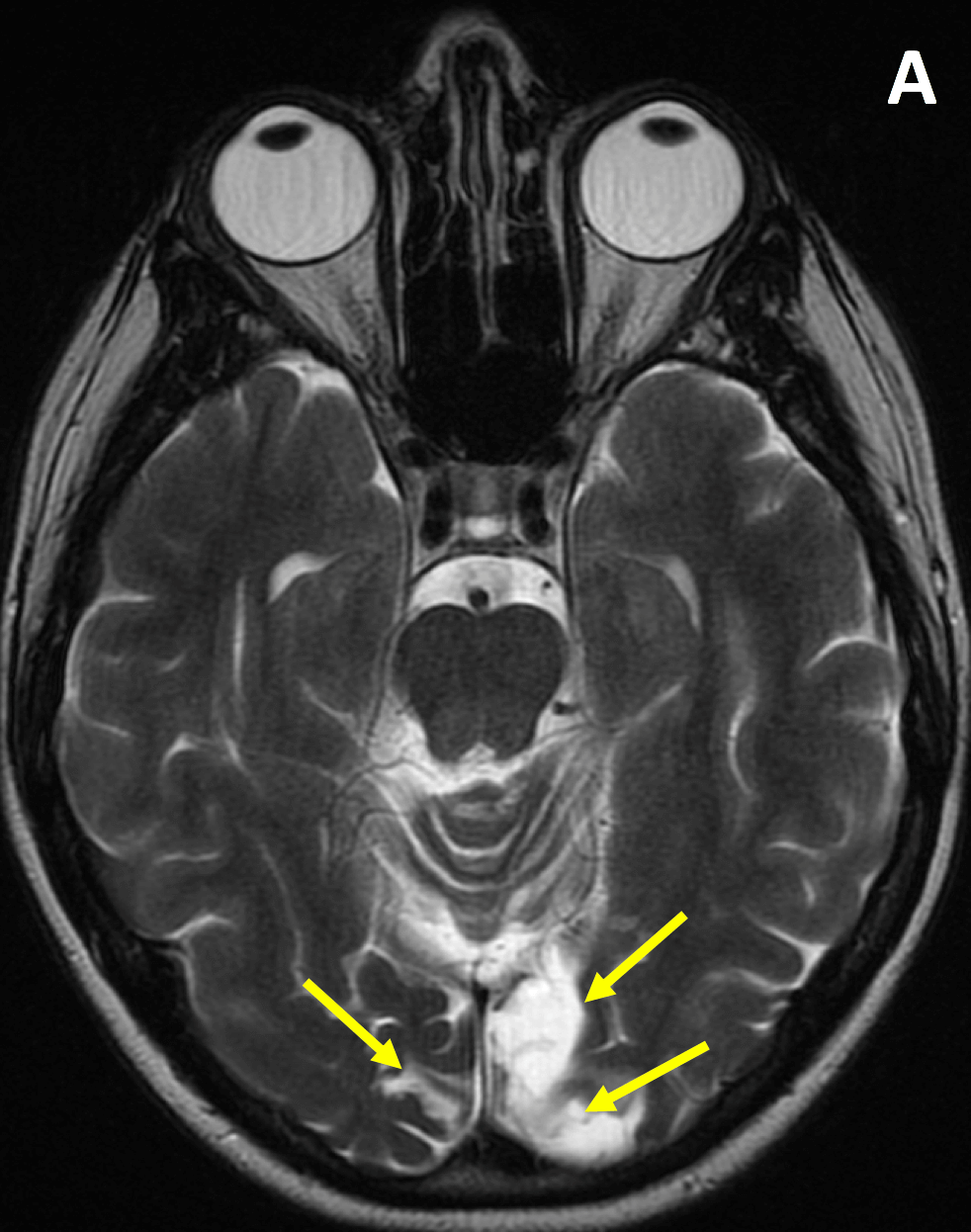
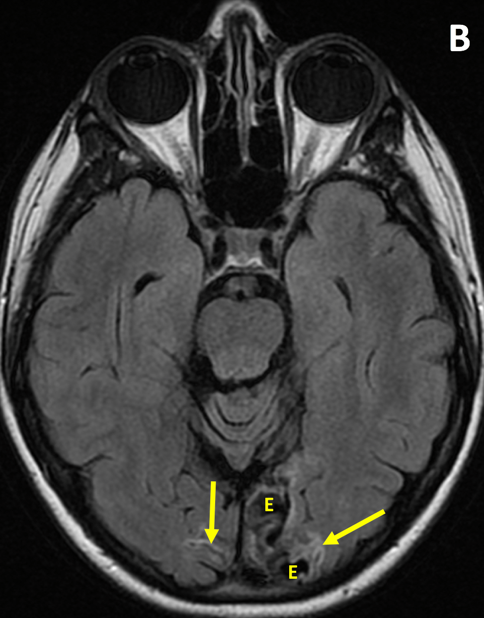
- Encephalomalacia and gliosis are high signal on T2-weighted images and often indistinguishable; on T2 FLAIR images, encephalomalacia is low signal and gliosis is high signal.
- The area/s of encephalomalacia should follow CSF on all pulse sequences (because it is a CSF filled cavity) and should demonstrate volume loss (as opposed to mass effect that would be seen with a space occupying cystic lesion)
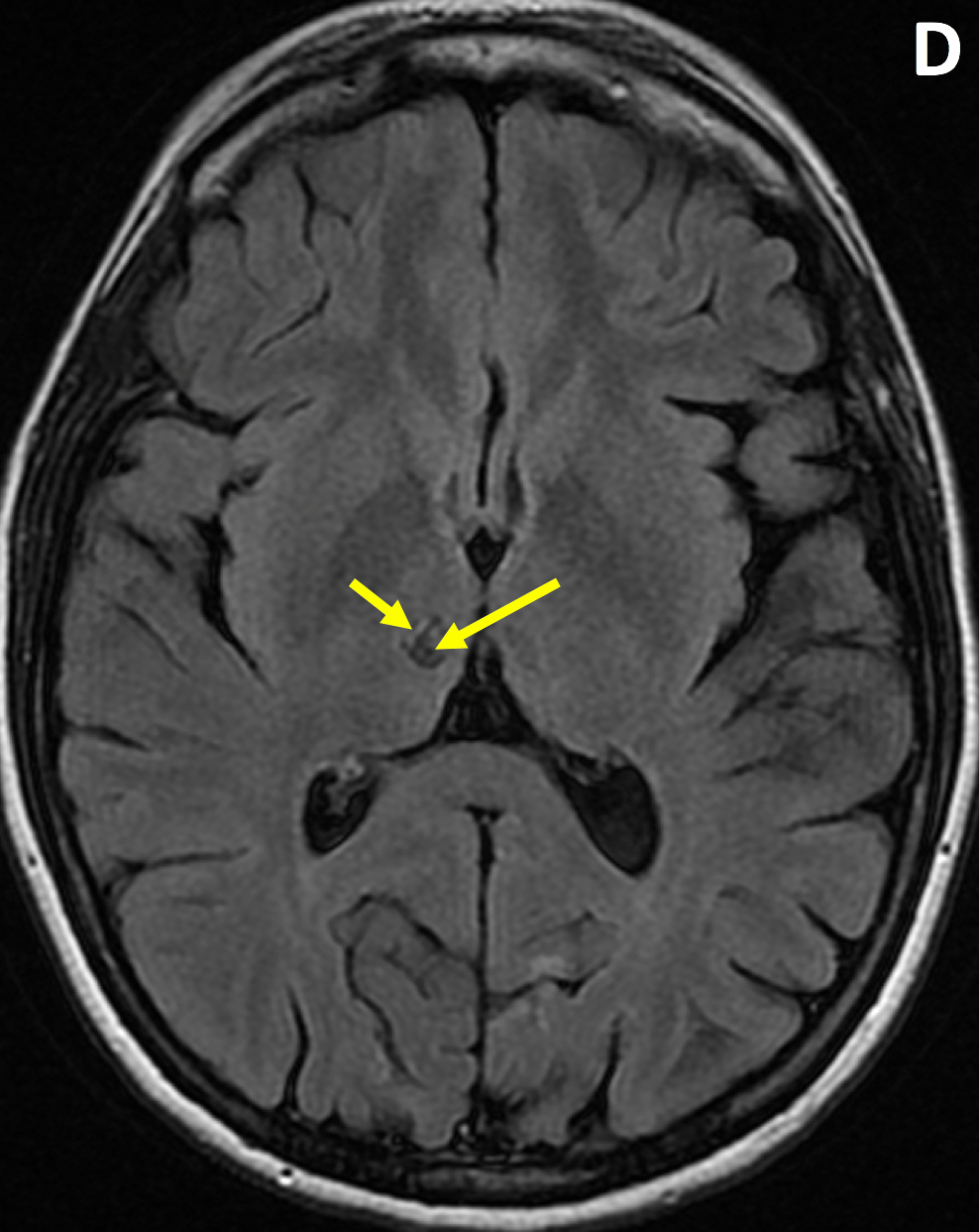
KEY IMAGES

Pearls
- A key finding in encephalomalacia is adjacent sulcal or ventricular enlargement with “negative” mass effect
- Areas of encephalomalacia can be asymptomatic, result in motor or sensory deficits, or represent a seizure focus
References
- Provenzale JM. Imaging of traumatic brain injury: a review of the recent medical literature. AJR AM J Roentgenol. 2010; 194(1):16-19
- Huang BY, Castillo M. Hypoxic-Ischemic Brain Injury: Imaging Findings from Birth to Adulthood. RadioGraphics 2008; 28:417-39
Case-based learning.
Perfected.
Learn from world renowned radiologists anytime, anywhere and practice on real, high-yield cases with Medality membership.
