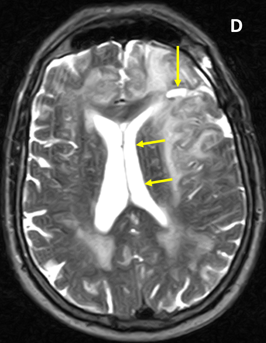
Diagnosis Definition
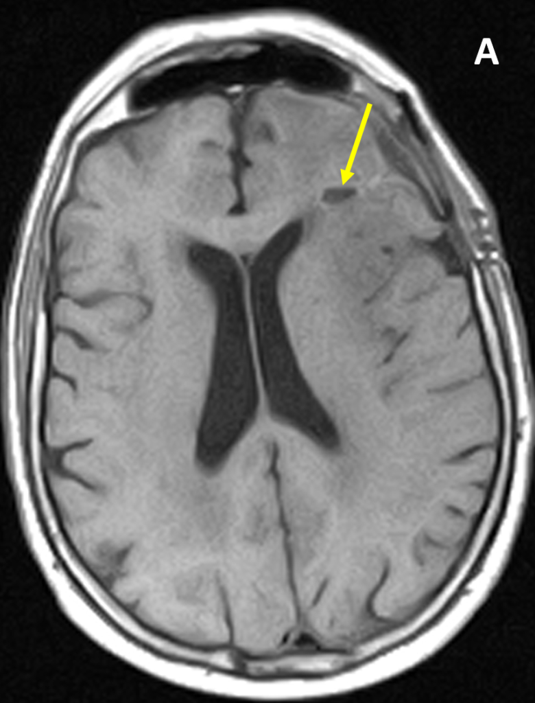
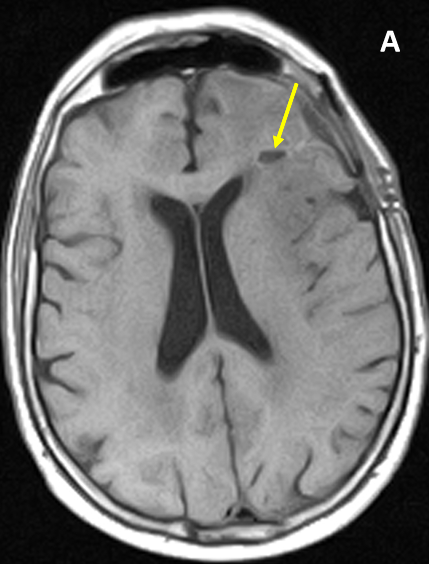
- Glioblastomas are classified by the World Health Organization (WHO) as grade IV malignant astrocytomas
- They are the most common type of adult primary brain tumor and have a poor prognosis, with a 10-20% 5-year survival
- Most occur in the supratentorial cerebral hemispheres, commonly involving the subcortical and periventricular white matter and crossing white matter tracts; they are occasionally multicentric
Imaging Findings
- CT of glioblastomas usually shows an avidly enhancing heterogeneous mass with hemorrhage and necrosis, significant mass effect, and marked surrounding vasogenic edema
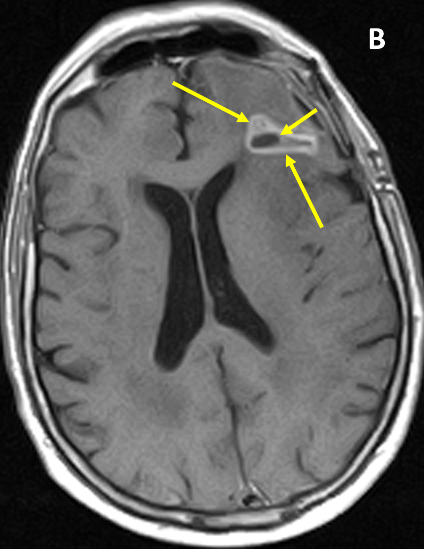
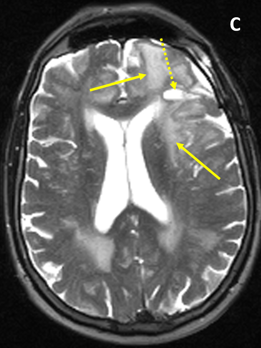
- MRI shows a poorly defined mass with mixed heterogeneous signal intensity (indicative of hemorrhage, necrosis, cyst formation, and contrast enhancement), mass effect, and surrounding edema
- Most high grade astrocytomas show restricted diffusion in the non-necrotic portion, have elevated perfusion, and reduced NAA/Choline ratios
- Thick irregular enhancement surrounding a central necrotic focus is the most common MRI finding and necrosis is the sina qua non of high grade astrocytomas
Pearls
- Involvement of both sides of the corpus callosum is described as a “butterfly” appearance
- The solid portions of glioblastoma may be densely cellular and show restricted diffusion
- Glioblastomas can infiltrate to other brain structures via the white matter tracts and disseminate along the ependyma and via the cerebrospinal fluid (CSF)
- The differential diagnosis of a glioblastoma includes other high grade neoplasms, CNS lymphoma, single large metastasis, and brain abscess
- MR spectroscopy and perfusion can help differentiate between a glioblastoma and a single large metastasis
References
- Okamoto K, Ito J, Takahashi N, et al. MRI of high-grade astrocytic tumors: early appearance and evolution. Neuroradiology 2002; 44:395–402
- Felix R, Schorner W, Laniado M, et al. Brain tumors: MR imaging with gadolinium-DTPA. Radiology 1985; 156:681-688
- Law M, Yang S, Wang H, et al. Glioma grading: sensitivity, specificity, and predictive values of perfusion MR imaging and proton MR spectroscopic imaging compared with conventional MR imaging. AJNR Am J Neuroradiol 2003; 24(10):1989-1998
Case-based learning.
Perfected.
Learn from world renowned radiologists anytime, anywhere and practice on real, high-yield cases with Medality membership.
- 100+ Mastery Series video courses
- 4,000+ High-yield cases with fully scrollable DICOMs
- 500+ Expert case reviews
- Unlimited CME & CPD hours

