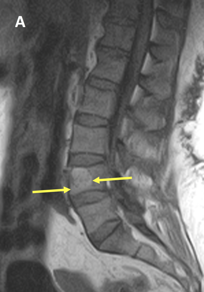
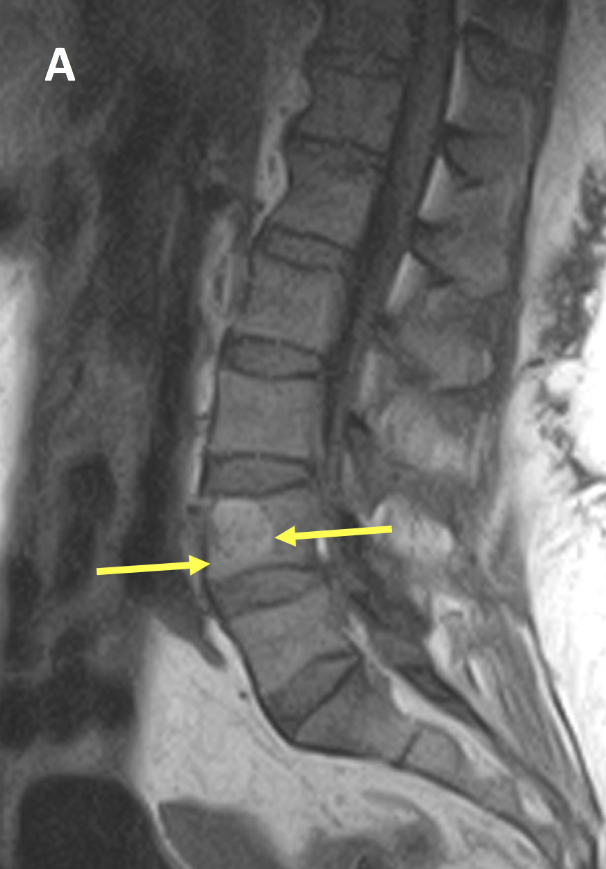
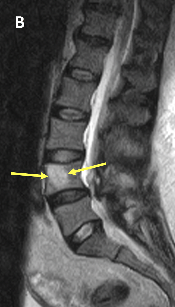
Diagnosis Definition
- Hemangiomas of the spine are venous vascular malformations
- Most are incidental findings and unrelated to patient symptoms
- When symptomatic, they can cause pain and myelopathy by intra-spinal bleeding, bony expansion or extra-osseous extension into surrounding soft tissue or the posterior neural elements
- Multiple hemangiomas are seen in the same patient in up to 60% of cases
- 10-12% of patients have vertebral hemangiomas at autopsy
Imaging Findings
- The classic CT appearance of a hemangioma is a well-circumscribed lesion with a sharp transition zone and sclerotic margin within the vertebral body; the lesion is hypoattenuating with a “polka-dot” pattern (due to a cross-section of reinforced trabeculae) or “corduroy” appearance (due to rarefaction with vertical striations) on axial images
- On MRI, they are typically hyperintense on T1 and T2-weighted images due to fat; the signal intensity varies on fat-saturated sequences depending on the amount of fat in the lesion relative to vascularity and interstitial edema
- Enhancement patterns are variable, but a mild degree of heterogeneous enhancement is common
Pearls
- Atypical hemangiomas may demonstrate hypointense signal on T1 and hyperintense signal on T2-weighted images and be difficult to distinguish from a malignant process; the morphology as seen on CT can often clarify the diagnosis
- The main differential diagnosis is focal fat in the vertebra, however, focal fat should not enhance whereas hemangiomas (even with fat in them) do; if the lesion is completely suppressed on a STIR sequence it is considered to be focal fat
- Hemangiomas are benign “do not touch” lesions, although if they abut the endplate or cortical bone, they can result in a pathologic fracture
References
- Ross JS, Masaryk TJ, Modic MT, et al. Vertebral hemangiomas: MR imaging. Radiology 1987; 165(1):165-169
- Baudrez V, Galant C, Vande Berg BC. Benign vertebral hemangioma: MR-histological correlation. Skeletal Radiol 2001; 30(8):442-446
Case-based learning.
Perfected.
Learn from world renowned radiologists anytime, anywhere and practice on real, high-yield cases with Medality membership.
- 100+ Mastery Series video courses
- 4,000+ High-yield cases with fully scrollable DICOMs
- 500+ Expert case reviews
- Unlimited CME & CPD hours


