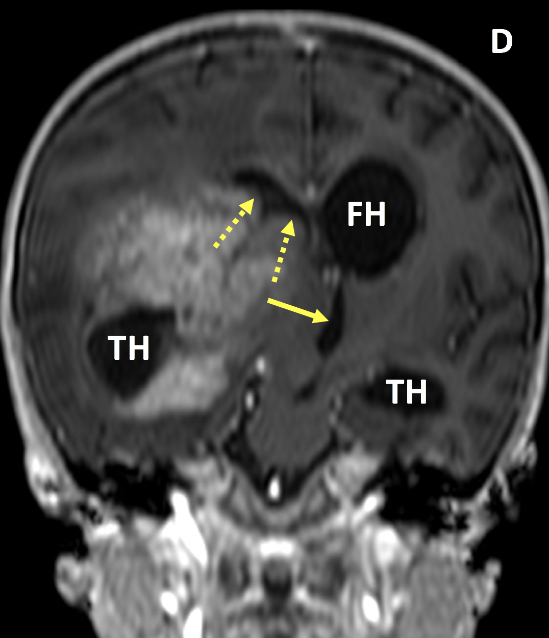
Diagnosis Definition
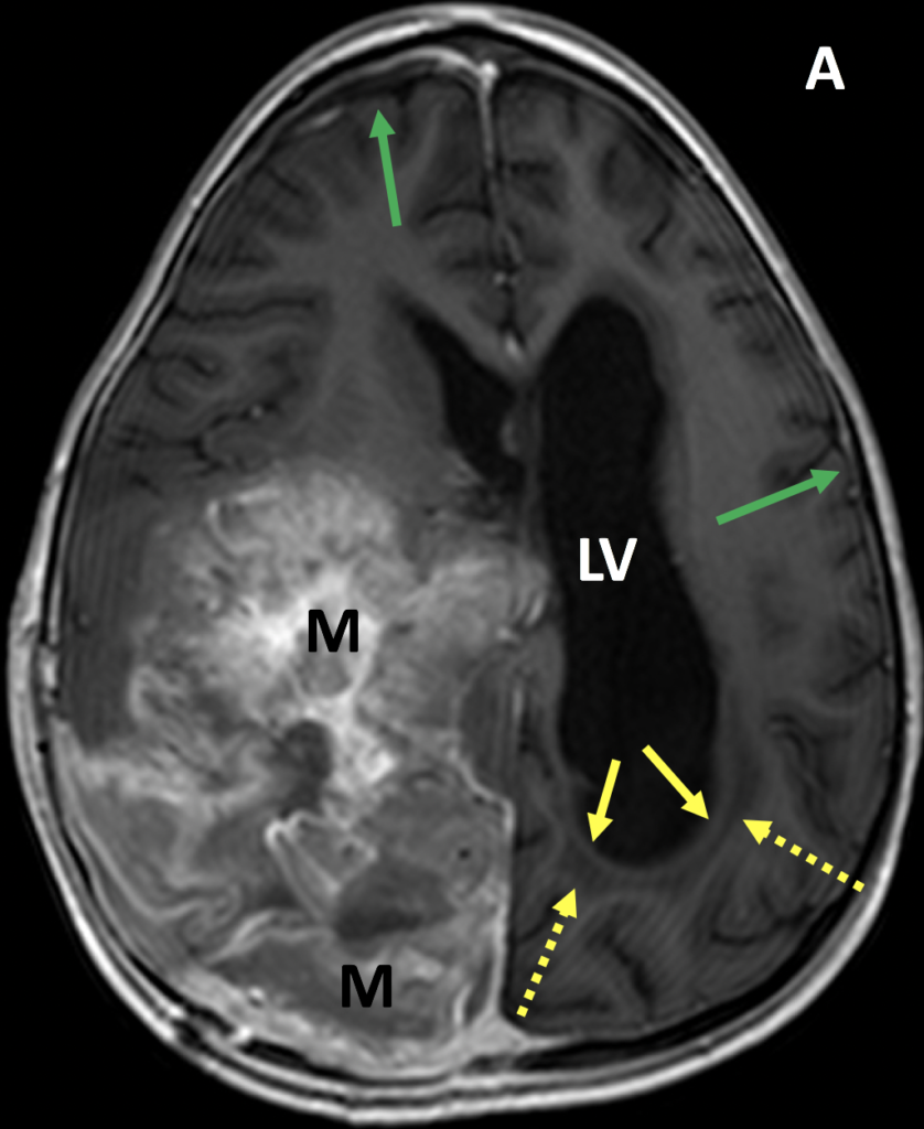
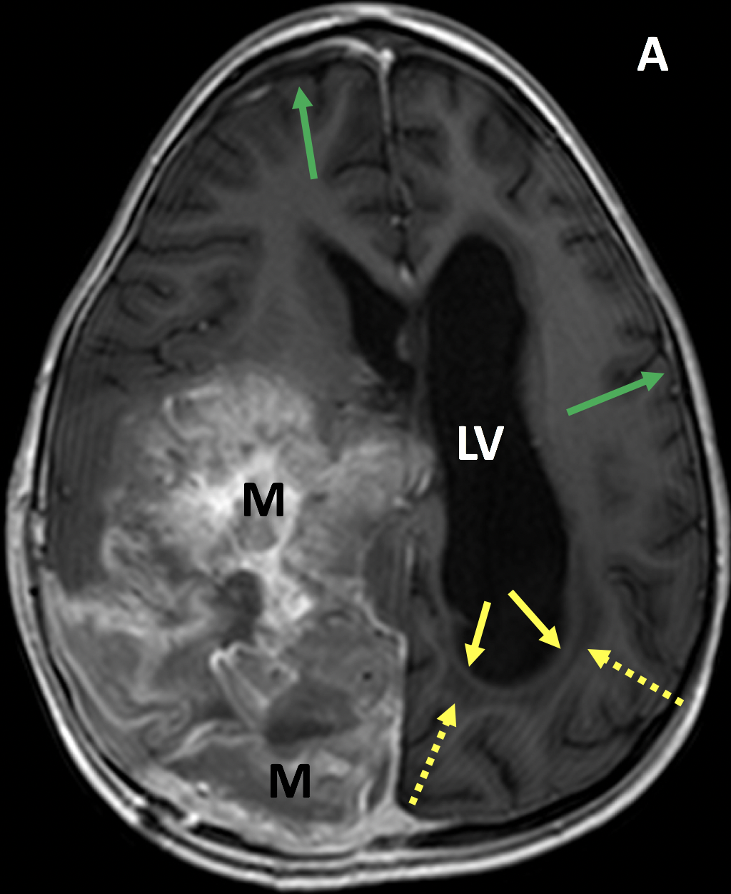
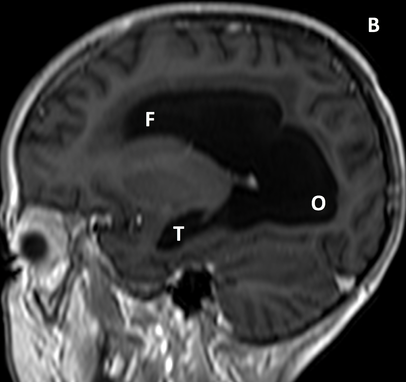
- Hydrocephalus (“water on the brain”) is excess cerebrospinal fluid (CSF) within the ventricles
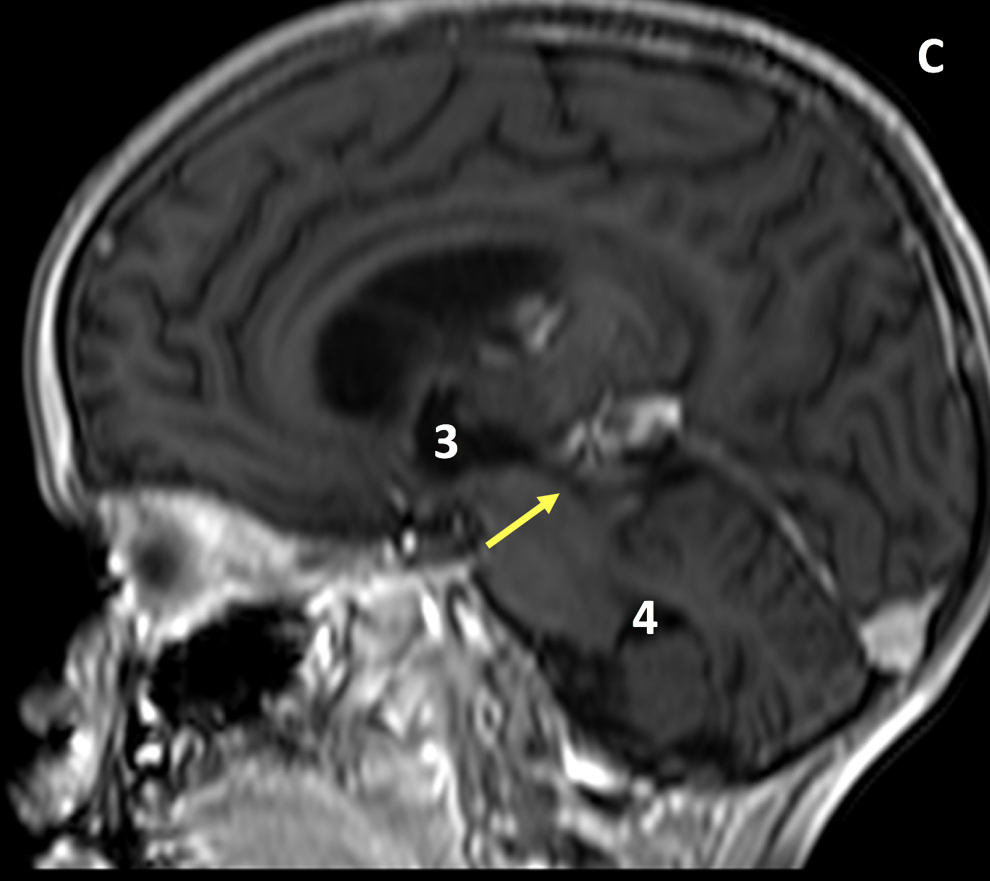
- CSF is secreted in the lateral ventricles, enters the 3rd ventricle through the foramen of Monro, flows into the 4th ventricle through the aqueduct of Sylvius, enters the subarachnoid space by the foramen of Magendie and foramina of Luschka, and is absorbed into the systemic venous system
- Causes of hydrocephalus include excessive CSF production, internal or external obstruction of CSF flow (obstructive hydrocephalus), or, more common in the elderly, impaired resorption of CSF (communicating hydrocephalus)
Imaging Findings
- CT and MRI of hydrocephalus show enlargement of the ventricles (generally or proximal to a point of obstruction) with excess CSF that follows fluid signal (low on T1 and high on T2)
- Ventricular enlargement can also reflect brain parenchymal volume loss (atrophy); features of hydrocephalus (distinct from atrophy) include an acute callosal angle, disproportionate prominence of the ventricles relative to the sulci, periventricular fluid representing transependymal CSF flow, rounding of the horns of the lateral ventricles, squaring of the gyri abutting the calvarium, partially empty sella, increased fluid along the optic nerves, and elevation of the optic disk
Pearls
- Since not all features of hydrocephalus are always present, the distinction between hydrocephalus and atrophy can be difficult
- Long-standing hydrocephalus itself can result in accelerated brain atrophy
- Normal pressure hydrocephalus (NPH), a type of communicating hydrocephalus, manifests with gait disturbance, urinary incontinence and dementia; early diagnosis and treatment with CSF diversion is critical
References
- Gibbs WN, Tanenbaum LN. Imaging of hydrocephalus. Appl Radiol 2018; 47(5):5-13
- Kartal MG, Algin O. Evaluation of hydrocephalus and other cerebrospinal fluid disorders with MRI: An update. Insights Imaging 2014; 5(4):531-541
Case-based learning.
Perfected.
Learn from world renowned radiologists anytime, anywhere and practice on real, high-yield cases with Medality membership.
- 100+ Mastery Series video courses
- 4,000+ High-yield cases with fully scrollable DICOMs
- 500+ Expert case reviews
- Unlimited CME & CPD hours

