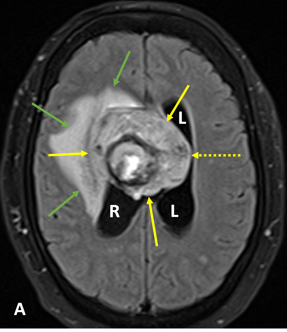
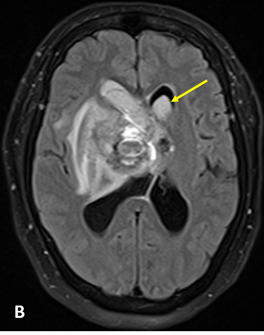
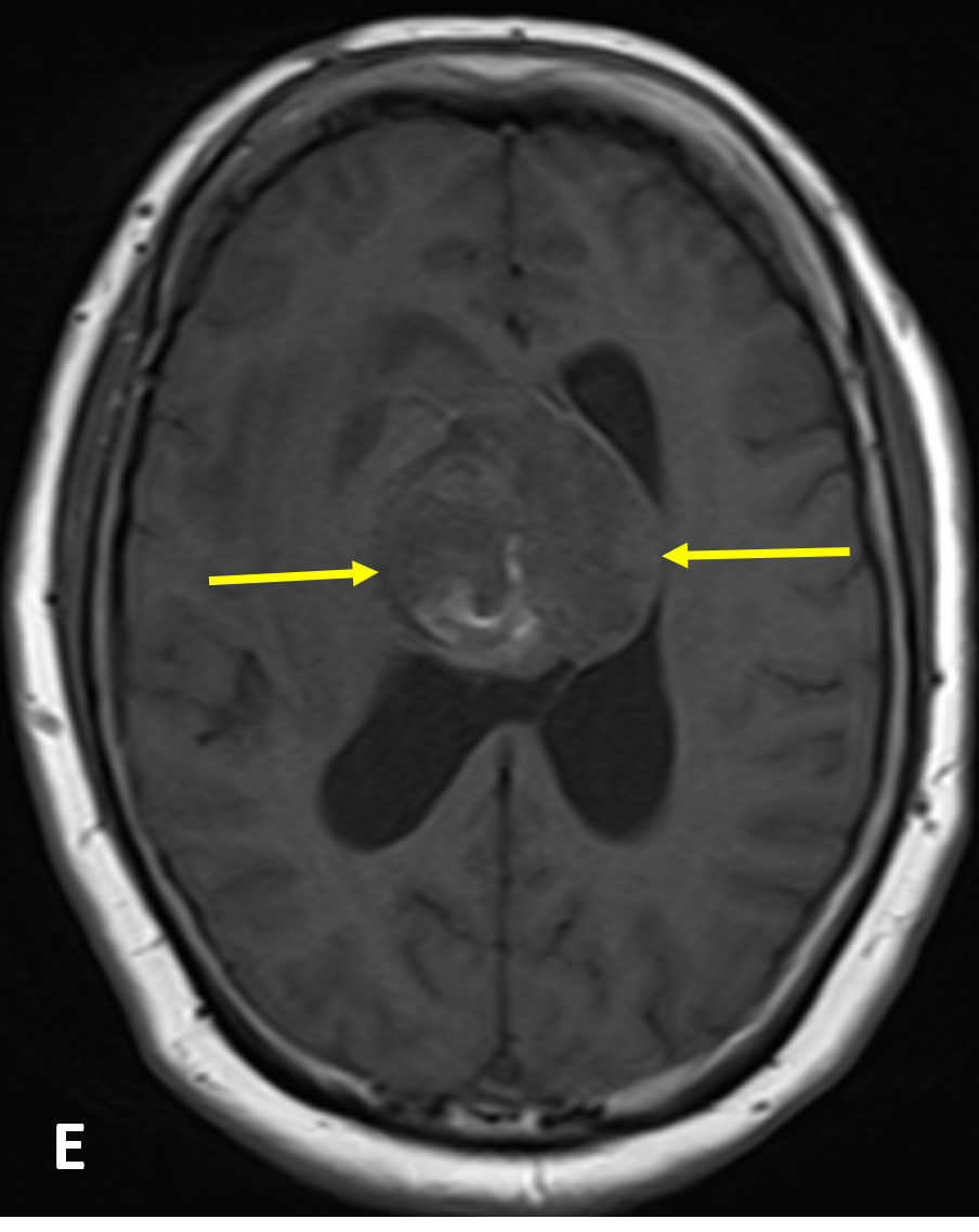
Intraventricular Hemorrhage (IVH)
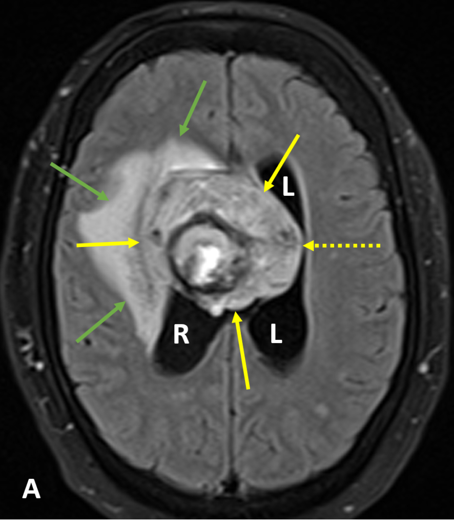
EXPLORE MORE DIAGNOSES EXPLORE MORE DIAGNOSES View Key Images View Key Images Review Stacks Review Stacks
  • Intraventricular hemorrhage (IVH) is defined as blood within the cerebral ventricular system, where cerebrospinal fluid (CSF) is produced and circulates through and towards the subarachnoid space
  • Most IVH is secondary and results from expansion of intraparenchymal or subarachnoid hemorrhage with extension into the ventricles
  • Primary IVH, all or largely confined to the ventricular space, is usually caused by trauma, aneurysm (associated with subarachnoid hemorrhage), vascular malformation, anticoagulation, or tumor (e.g. choroid plexus, ependymoma, metastases)
  • IVH is a distinct entity in newborns
  • Non-contrast CT scans show hyperintense blood in the ventricles, tending to pool dependently, best seen in the occipital horns; acutely, the blood can clot and form a ‘cast’
  • MRI is more sensitive than CT in showing small amounts of blood, especially in the posterior fossa, and old blood products such as hemosiderin and hemosiderosis staining the ependyma
  • Both FLAIR and susceptibility weighted imaging (SWI) sequences are particularly sensitive to small amounts of blood
  • Within 48 hours blood will appear as hyperintense to the attenuated adjacent CSF on FLAIR images; after this time the signal is more variable and can be difficult to distinguish from flow related artifact unless other sequences are also used
  • IVH is associated with significant morbidity when due to intracerebral hypertensive hemorrhage
  • IVH can lead to a clot that blocks flow and leads to obstructive hydrocephalus, which may quickly result in increased intracranial pressure and death
  • Breakdown products from an intraventricular clot may generate an inflammatory response that damages the arachnoid granulations, inhibiting the regular reabsorption of CSF and resulting in permanent communicating hydrocephalus
  • Isolated blood in the occipital horns of a trauma patient implies a tear in the splenium of the corpus callosum, well depicted on FLAIR and SWI sequences

1.  Sohn CH, Baik SK, Lee HJ et al. MR imaging of hyperacute subarachnoid and intraventricular hemorrhage at 3T: a preliminary report of gradient echo T2*-weighted sequences. AJNR Am J Neuroradiol 2005; 26(3): 662-5

2.  Bakshi R, Kamran S, Kinkel PR et al. Fluid-attenuated inversion-recovery MR imaging in acute and subacute cerebral intraventricular hemorrhage. AJNR Am J Neuroradiol 1999; 20(4): 629-36

Case-based learning.
Perfected.

Try MRI Online Premium for free.


100+

Mastery Series video courses

3000+

High-yield cases

500+

Expert case reviews

Unlimited

CME & SA-CME credits

Learn from world renowned radiologists anytime,
actice on real, high-yield cases with MRI Online Premium.