Diagnosis Definition
- The Lisfranc joint complex includes the tarsometatarsal (TMT), intermetatarsal (IMT), and intertarsal joints
- The Lisfranc ligament attaches the medial cuneiform to the base of the 2nd MT and has 1) dorsal, 2) interosseous, and 3) plantar components; variable ligaments attach 2nd-5th MT bases and the cuneiforms to their neighbors
- Lisfranc injuries range from sprain to fracture with or without dislocation and result from crushing or rotational force on a plantar flexed forefoot
- Fractures are classified as 1) homolateral (MTs displaced in same direction), 2) isolated (1-2 MTs displaced) or 3) divergent (MTs displaced in opposite directions)
Imaging Findings
- MRI sequences include sagittal T1 and STIR, axial PD and T2 Fat-sat (FS), and coronal PD and T2 FS
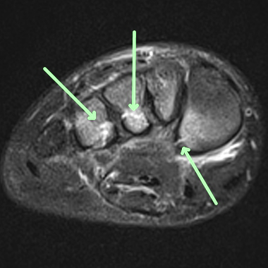
- The normal Lisfranc ligament is homogeneous or striated and has low or intermediate signal
- On MRI, Lisfranc injuries may be seen as high signal bone bruises, fractures, dislocations, increased signal within the ligaments, periligamentous edema, or ligament disruption
KEY IMAGES

Pearls
- Undiagnosed Lisfranc sprains can lead to chronic instability and early osteoarthritis
- Low signal scars of chronic ligament rupture may simulate an intact ligament on T2
- Tarsometatarsal edema and multiple microfractures are highly suggestive of Lisfranc injury
References
- Cheung Y, Rosenberg ZS. MR imaging of ligamentous abnormalities of the ankle and foot. MRI Clinics of North America 2001; 9:507-31
Case-based learning.
Perfected.
Learn from world renowned radiologists anytime, anywhere and practice on real, high-yield cases with Medality membership.
