Diagnosis Definition
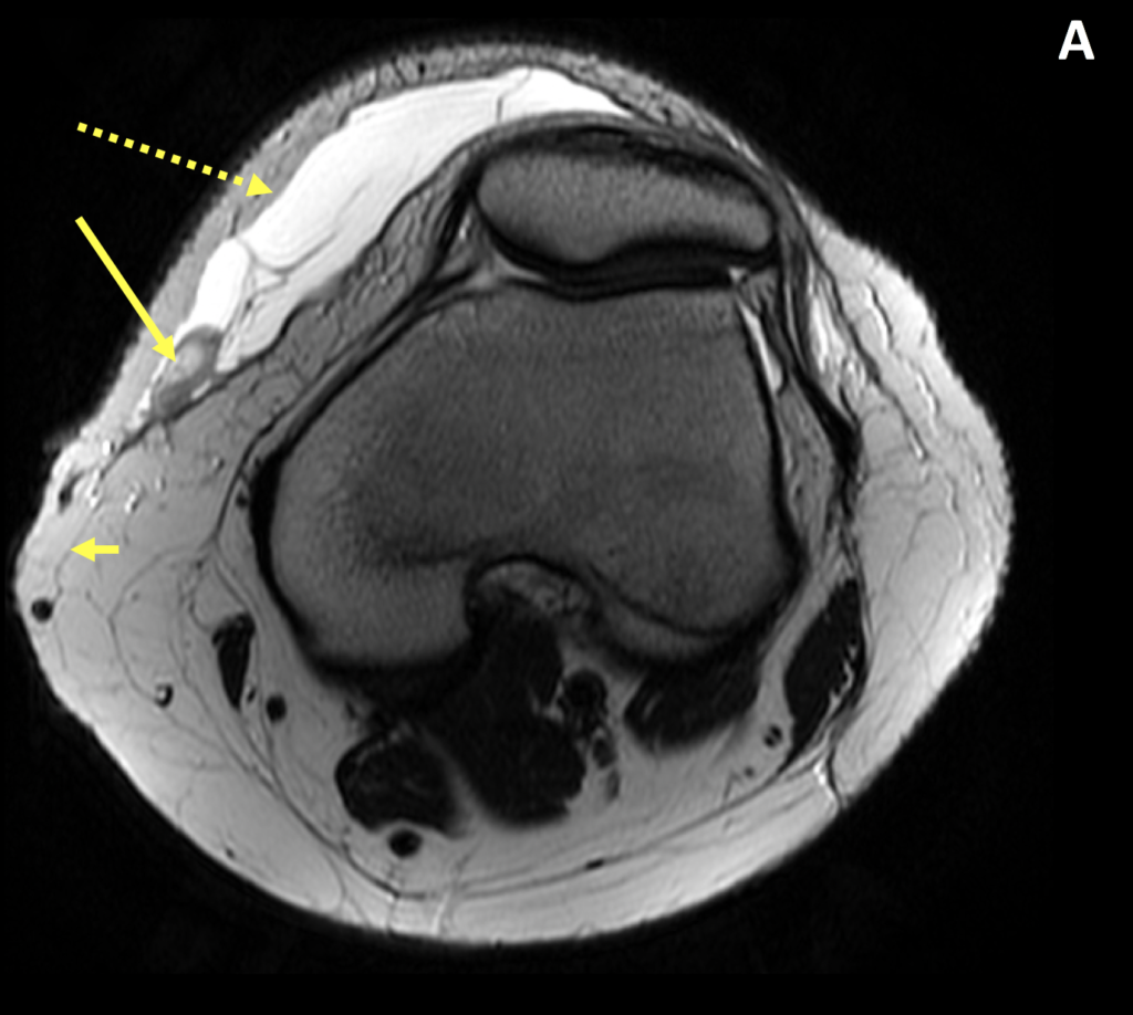
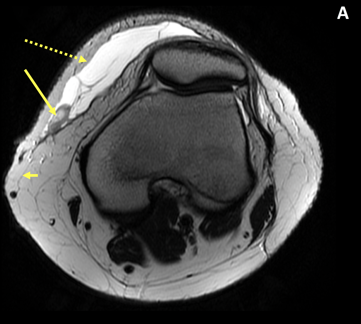
- The Morel-Lavallee lesion (MLL) results from a closed traumatic degloving soft tissue injury that most commonly occurs in the area of the greater trochanter and knee
- The injury results in separation of the hypodermis from the underlying fascia when a shearing force is applied to the soft tissue, disrupting the perforating vascular and lymphatic structures
- A mixture of blood, lymph, and fatty debris collects in the potential space, which is replaced by serosanguinous fluid as the lesion enlarges; if left untreated, inflammation can lead to pseudocapsule formation
- Treatment options include percutaneous drainage with sclerodesis, however, once collections are encapsulated, surgical drainage and resection may be required
Imaging Findings
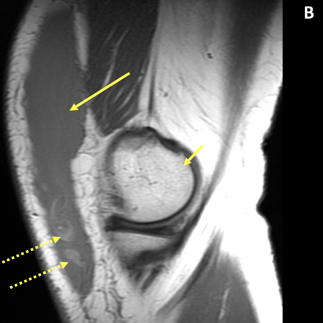
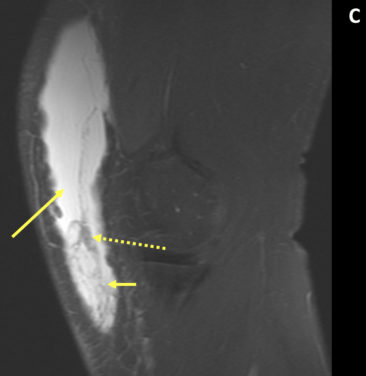
- On MRI, the MLLs may be of variable signal intensity depending on the age and constituents of the fluid collection
- Fibrinous and hemorrhagic debris appears hypointense on T2-weighted images and fat globules appear as dependently positioned hyperintense areas on T1-weighted images
- Fluid-fluid levels and septations may be seen
- When a pseudocapsule forms, the collections become circumscribed
Pearls
- Fat globules are a specific feature of an MLL
- Infection, pseudocyst formation, and cosmetic deformity can result from improper or untimely diagnosis and management
- Most common location is the hip followed by the knee
References
- Gilbert BC et al. MRI of a Morel-Lavallée Lesion. AJR 2004; 182(5):1347-1348
- Vassalou EE et al. Morel-Lavallée Lesions of the Knee: MRI Findings Compared With Cadaveric. AJR 2018; 210(5):234-239
- Chew FS. Imaging of Soft Tissue Lesions and Calcifications. In: Musculoskeletal Imaging: The Essentials. Wolter Kluwer; 2001:261-262
Case-based learning.
Perfected.
Learn from world renowned radiologists anytime, anywhere and practice on real, high-yield cases with Medality membership.
- 100+ Mastery Series video courses
- 4,000+ High-yield cases with fully scrollable DICOMs
- 500+ Expert case reviews
- Unlimited CME & CPD hours


