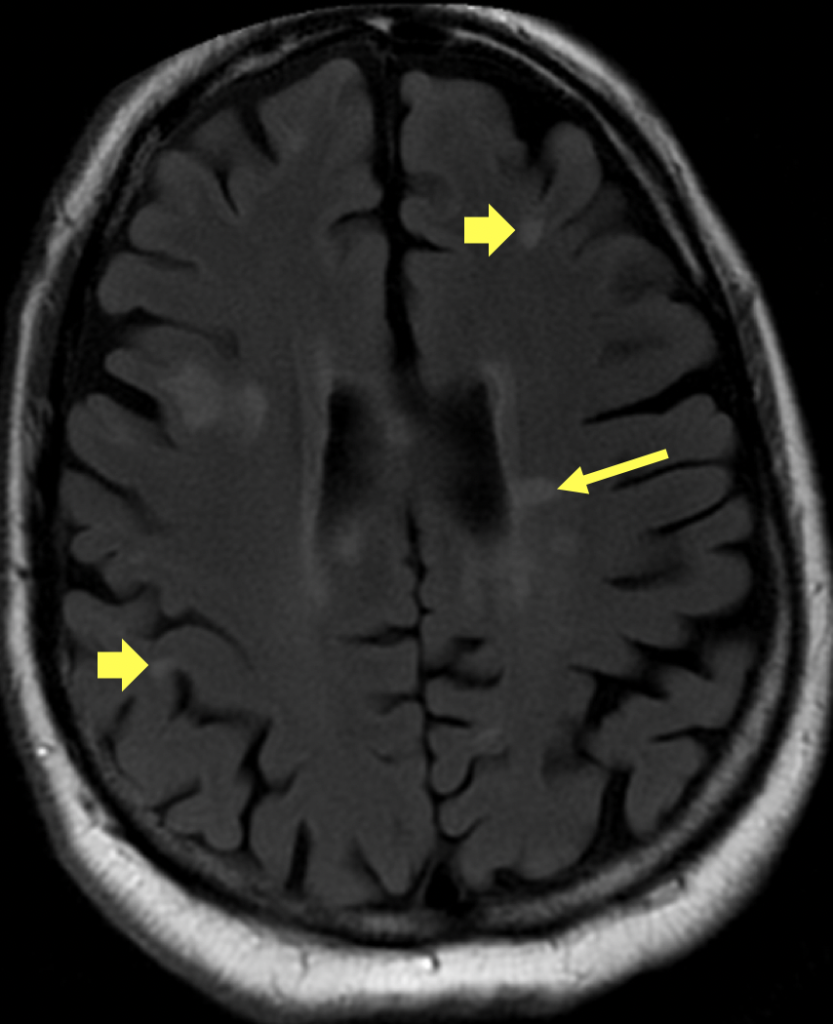
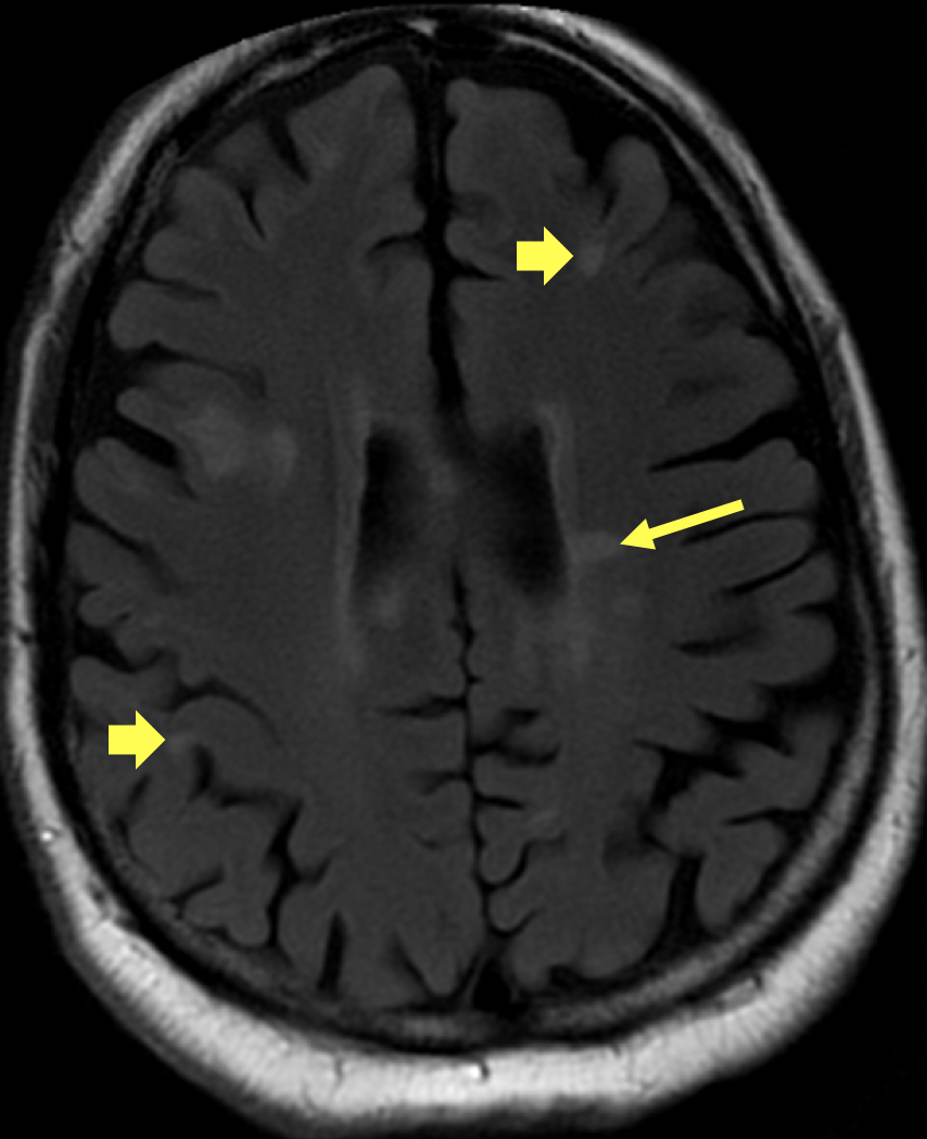
Diagnosis Definition
- MS is defined histopathologically as inflammatory demyelination in the central nervous system (CNS); the etiology is unknown
- The age of onset is usually between 15-50 (average age 34) with a 2:1 female predominance
- MS is most commonly relapsing-remitting (i.e., clearly defined relapses with no disease progression between them)
Imaging Findings
- The “McDonald diagnostic criteria” includes dissemination of lesions in both space (one or more hyperintense lesions on T2-weighted imaging in two of the following areas: periventricular, juxtacortical, infratentorial, and spinal cord) and time (new T2 hyperintense lesion on follow-up MRI or simultaneous presence of enhancing and nonenhancing lesions on a single MRI)
- Lesions are usually ovoid, well demarcated, and oriented perpendicular to the margins of the lateral ventricles
- Nodular and peripheral enhancement of lesions on post-contrast T1-weighted images, or diffusion restriction, indicates active demyelination
- Tumefactive demyelination can present as larger masslike lesions
Pearls
- Serial MRI examinations are used to monitor disease course and guide clinical management, including changes in therapy
- Initial periventricular lesions may be linear (aka, “Dawson fingers”), distinguishing MS from other white matter disorders
- T2-weighted non-FLAIR images are often superior to FLAIR images for showing infratentorial (posterior fossa) lesions
- Incomplete peripheral lesion enhancement (aka, “open ring” sign) can distinguish demyelination from other processes
- The mass effect and edema associated with tumefactive demyelination is disproportionately less than expected for the size of the lesion, helping to differentiate it from neoplasm
- Perfusion weighted MR images show tumefactive demyelinating lesions to have low perfusion whereas high grade astrocytomas are hyperperfused
References
- Vigeveno RM, Wiebenga OT, Wattjes MP, et al. Shifting imaging targets in multiple sclerosis: from inflammation to neurodegeneration. J Magn Reson Imaging 2012; 36(1):1-19
- Polman CH, Reingold SC, Banwell B, et al. Diagnostic criteria for multiple sclerosis: 2010 Revisions to the McDonald Criteria. Ann Neurol 2011; 69:292-302
Case-based learning.
Perfected.
Learn from world renowned radiologists anytime, anywhere and practice on real, high-yield cases with Medality membership.
- 100+ Mastery Series video courses
- 4,000+ High-yield cases with fully scrollable DICOMs
- 500+ Expert case reviews
- Unlimited CME & CPD hours


