Diagnosis Definition
- Pituitary adenomas are benign neoplasms affecting individuals of all ages, peaking between the third and sixth decades of life
- They represent the most common sellar tumor; those exceeding 10 mm are defined as macroadenomas (but most are microadenomas)
- Patients may be asymptomatic or may present with symptoms related to hormonal imbalance or mass effects; such hormones include prolactin, thyrotropin, adrenocorticotropin, cortisol, luteinizing hormone follicle-stimulating hormone, growth hormone, insulinlike growth factor-1 (IGF-1), and alpha subunit glycoprotein
Imaging Findings
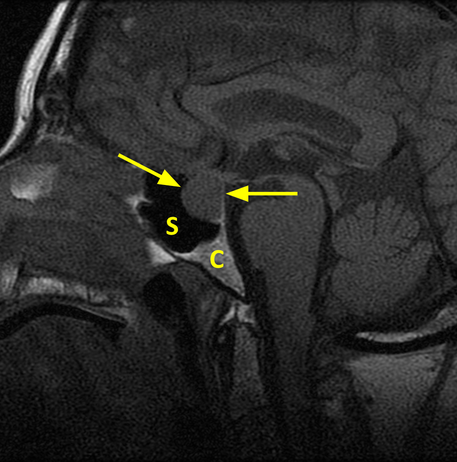
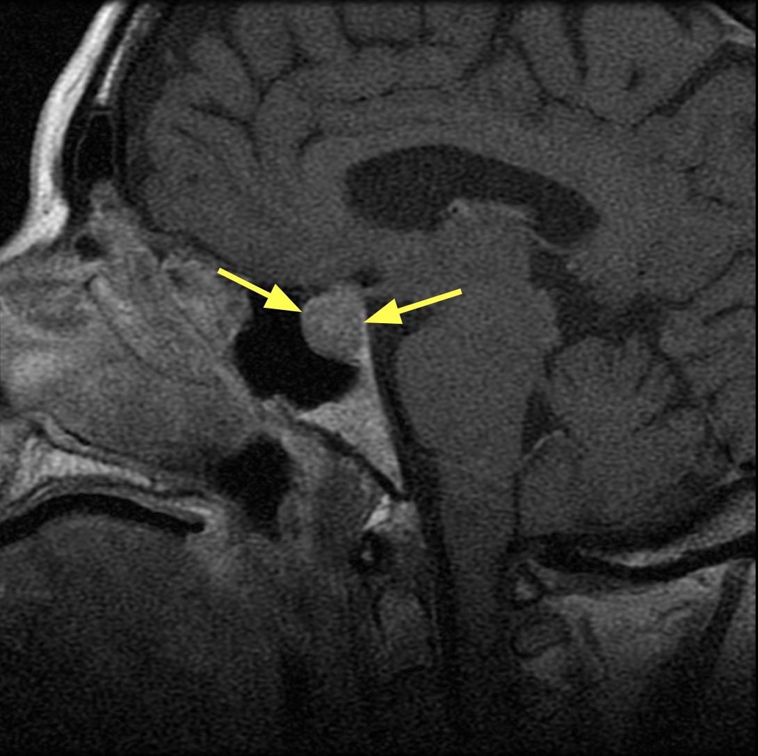
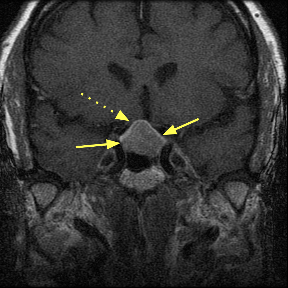
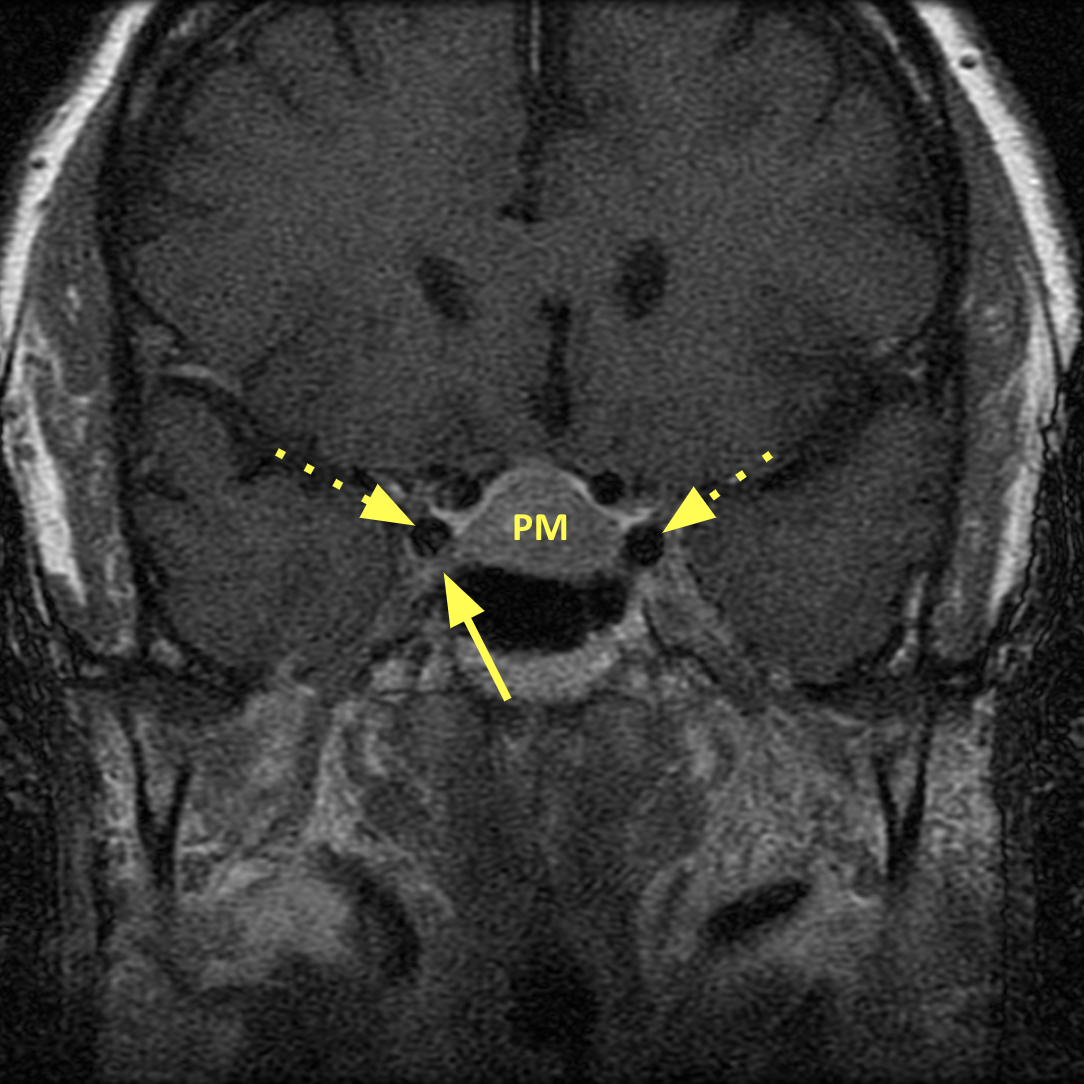
- Pituitary adenomas are seen on CT and MRI as abnormal tissue in the sella and absence of a normal appearing pituitary gland
- Most microadenomas are seen as low enhancing hypointense defects within an enhancing gland and are best detected on dynamic post contrast T1 weighted images
- Most adenomas are NOT avidly enhancing; they often enhance the same as the normal pituitary or slightly less, rarely more than the normal pituitary
- The sella is typically enlarged and there may be invasion of the skull base; there is often mass effect superiorly on the optic chiasm or growth laterally into the cavernous sinus; less commonly seen is downward growth and invasion of the sellar floor, sphenoid sinus and clivus
Pearls
- Differential diagnoses of tumors with calcification, such as germinomas, craniopharyngiomas, and meningiomas, are better determined with CT scanning
- Visual field testing should be performed, especially in tumors involving the optic chiasm
- Pituitary apoplexy, which tends to occur in macroadenomas, results from infarction of a pituitary tumor or sudden hemorrhage within; this presents as a medical emergency with a headache, sudden collapse, shock, and death if not treated emergently (note: pituitary apoplexy can also occur in the absence of an adenoma)
References
- Pisaneschi M, Kapoor G. Imaging the sella and parasellar region. Neuroimaging Clin N Am 2005;15:203–19
- Hess CP, Dillon WP. Imaging the pituitary and parasellar region. Neurosurg Clin N Am 2012; 23:529–42
- Chin BM, Orlandi RR, Wiggins III RH. Evaluation of the sellar and parasellar regions. Magn Reson Imaging Clin N Am 2012; 20:515-543
Case-based learning.
Perfected.
Learn from world renowned radiologists anytime, anywhere and practice on real, high-yield cases with Medality membership.
- 100+ Mastery Series video courses
- 4,000+ High-yield cases with fully scrollable DICOMs
- 500+ Expert case reviews
- Unlimited CME & CPD hours


