Diagnosis Definition
- Sacral insufficiency fractures (SIFs) result from normal stress applied to abnormal bone; antecedent trauma is not identified in two-thirds of patients
- Patients most commonly present with diffuse low back pain, which may radiate to the buttock, hip, or groin
- There is often a delay in diagnosis because clinical symptoms are frequently vague and nonspecific and can mimic a variety of pathologic processes, including radiculopathy and metastatic disease
- SIFs commonly affect elderly women with osteoporosis; other risk factors include pelvic radiation, steroid-induced osteopenia, rheumatoid arthritis, multiple myeloma, Paget disease, renal osteodystrophy, and hyperparathyroidism
- Almost all patients with SIFs are older than 55, with a mean age between 70 and 75 years
- SIFs are unilateral or bilateral with equal frequency
Imaging Findings
- The sacrum is divided into Zone 1 (sacral ala and bone between the sacroiliac joints and the neural foramina; the most common site of SIFs), Zone 2 (neural foramina), and Zone 3 (sacral bodies)
- A characteristic “H” sign is seen on bone scintigraphy
- CT scans show sclerosis in the sacral ala and lateral sacrum paralleling the SI joints, vertical fracture lines on axial images, and full fracture lines on coronal reconstructions
- MR imaging can detect early changes of sacral insufficiency and, similar to bone scintigraphy, has a reported sensitivity at or near 100%
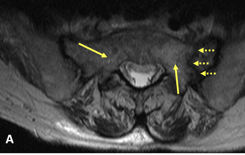
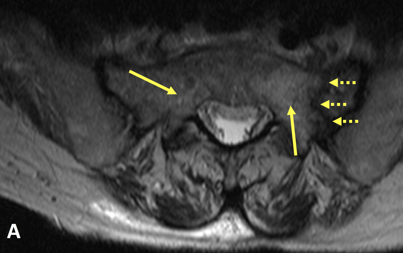
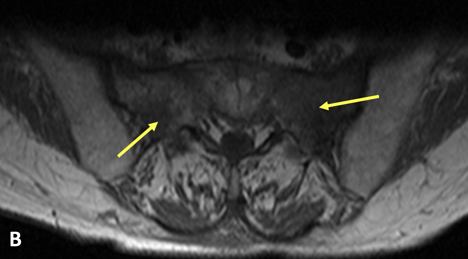
- MR T2 STIR sequences are exquisitely sensitive for the detection of early marrow edema related to SIFs, which can be seen as early as 18 days after symptoms develop
- MRI shows marrow edema (hyperintensity on T2-weighted and STIR images and low signal on T1) and a hypointense fracture line in the area of edema
Pearls
- There is a high incidence of concomitant pelvic insufficiency fractures, especially of the pubic rami and parasymphyseal region
- Neurologic symptoms related to SIFs are unusual
References
- Lyders EM, Whitlow CT, Baker MD, Morris PP. Imaging and Treatment of Sacral Insufficiency Fractures. American Journal of Neuroradiology 2010; 31(2):201-210
- Denis F, Davis S, Comfort T. Sacral fractures: an important problem. Clin Orthop Relat Res 1988; 227:67-81
Case-based learning.
Perfected.
Learn from world renowned radiologists anytime, anywhere and practice on real, high-yield cases with Medality membership.
- 100+ Mastery Series video courses
- 4,000+ High-yield cases with fully scrollable DICOMs
- 500+ Expert case reviews
- Unlimited CME & CPD hours


