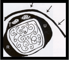
Diagnosis Definition
- The ulnar collateral ligament (UCL) of the thumb arises from the dorsal MC head and inserts at the base of the proximal phalanx
- Most UCL injuries occur from a fall on an abducted, outstretched thumb, producing a valgus force on the MCP joint
- Stener lesions occur in 64-88% of complete UCL tears; the distal portion of the UCL avulses from its attachment on the proximal phalanx and herniates through the adductor aponeurosis
- Because the torn end of the UCL cannot approximate the proximal phalangeal insertion, normal healing and stability cannot be achieved and surgery is required to repair the UCL
Imaging Findings
- Key MRI sequence is coronal oblique with T2 FSE
- On fluid-sensitive sequences, partial tears of the UCL demonstrate focal attenuation of the ligament with hyperintense signal partially traversing the ligament
- Complete tears are identified by hyperintense signal fully traversing the ligament or at the site of bony attachment on fluid-sensitive sequences
- MRI of a Stener lesion shows a “yo-yo on a string” appearance with the string representing the adductor aponeurosis and the yo-yo representing the balled-up and proximally retracted UCL
Pearls
- Simply following the adductor aponeurosis proximally on the axial images provides a useful landmark for identifying displaced ligamentous tissue, which is readily seen protruding beyond the plane of the aponeurosis
- Gamekeeper’s thumb (chronic injury) and skier’s thumb (acute injury) are two similar conditions involving the UCL
References
- Hergan K, Mittler C, Oser W. Ulnar collateral ligament: differentiation of displaced and nondisplaced tears with US and MR imaging. Radiology 1995; 194(1):65-71
Case-based learning.
Perfected.
Learn from world renowned radiologists anytime, anywhere and practice on real, high-yield cases with Medality membership.
- 100+ Mastery Series video courses
- 4,000+ High-yield cases with fully scrollable DICOMs
- 500+ Expert case reviews
- Unlimited CME & CPD hours


