Diagnosis Definition
- TSC is an autosomal dominant, multisystem disease characterized by hamartomas in multiple organs
- The classic triad of symptoms (seizures, mental retardation, and adenoma sebaceum) is seen in less than 30% of cases
- Seizures develop in the majority of patients
- A definitive diagnosis includes either 2 major features or 1 major and 2 minor features from a list of skin, intracranial, retinal, cardiac, renal, rectal, bone, dental, lymphatic, and gingival features
Imaging Findings
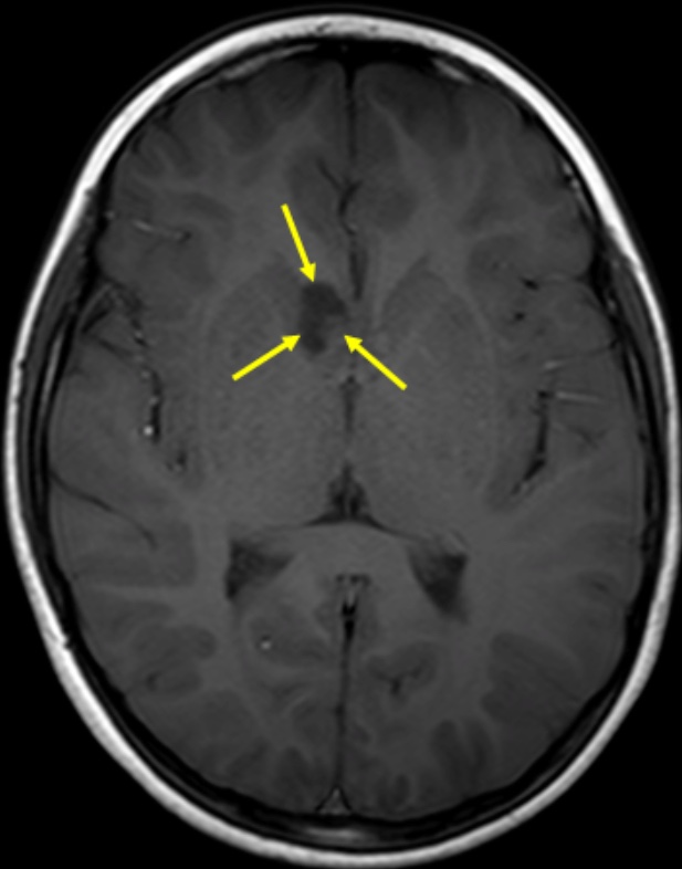
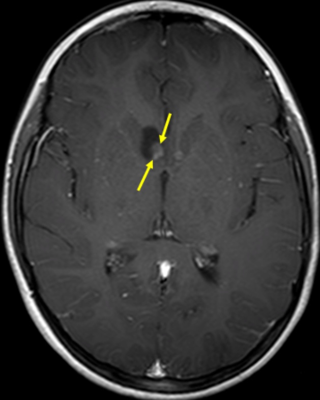
- In 90% of patients with TSC, MRI shows rounded, benign lesions (subependymal nodules) along the walls of the lateral ventricles, commonly near the foramen of Monro; the nodules are isointense to mature white matter, can enhance with contrast, and often calcify
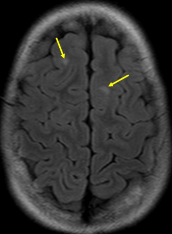
- Another common feature of TSC is multiple epileptogenic cortical/subcortical hamartomas (or tubers), which manifest most commonly in the frontal lobes as expanded gyri; they appear as low signal areas on T1-weighted images and high signal areas on T2-weighted and FLAIR images
- Subependymal nodules can degenerate into subependymal giant cell astrocytomas (SEGAs) in up to 15% of patients with TSC; they are most commonly seen near the foramen of Monro, often have cystic and solid components that partially enhance, and can grow large enough to cause ventricular obstruction and hydrocephalus
Pearls
- The critical imaging finding that differentiates SEGAs from subependymal nodules is that SEGAs tend to enlarge; also, SEGAs enhance and most subependymal nodules do not
- Hyperintense linear or curvilinear streaks extending from the ventricle to the cortex on T2-weighted images are thought to represent “migration lines” or bands of hypomyelinated disordered cells
- Tubers often calcify, which is best appreciated on CT scans
References
- Kalantari B, Salamon N. Neuroimaging of tuberous sclerosis: spectrum of pathologic findings and frontiers in imaging. AJR 2008; 190(5):304-309
- DiPaolo D, Zimmerman RA. Solitary cortical tubers. Am J Neuroradiol 1995; 16:1360-1364
Case-based learning.
Perfected.
Learn from world renowned radiologists anytime, anywhere and practice on real, high-yield cases with Medality membership.
- 100+ Mastery Series video courses
- 4,000+ High-yield cases with fully scrollable DICOMs
- 500+ Expert case reviews
- Unlimited CME & CPD hours


